. 2011 Apr 15;117(8):1630-9.
doi: 10.1002/cncr.25724. Epub 2010 Nov 8.

MicroRNA profiling of adrenocortical tumors reveals miR-483 as a marker of malignancy

Affiliations

MicroRNA profiling of adrenocortical tumors reveals miR-483 as a marker of malignancy

Erin E Patterson et al. Cancer. .

Abstract

Background: The authors are interested in identifying molecular markers that can aid in the diagnosis of adrenocortical carcinoma (ACC). The aim of this study was to identify microRNAs (miRNAs or miRs) that are differentially expressed in malignant adrenocortical tumors as compared with benign tumors and assess their potential as diagnostic predictors.

Methods: Differentially expressed miRNAs were identified using microarray profiling of adrenocortical tumors and validated by quantitative real-time RT-PCR.

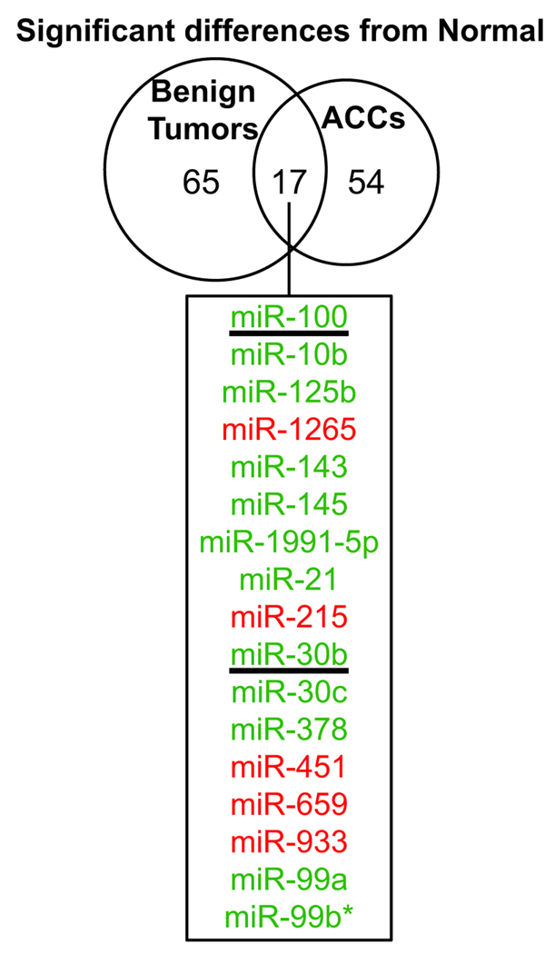
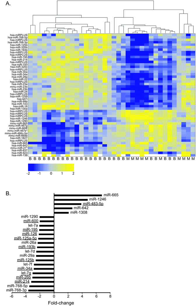
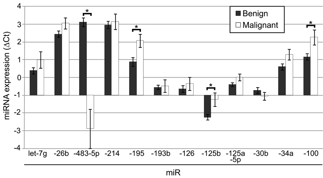
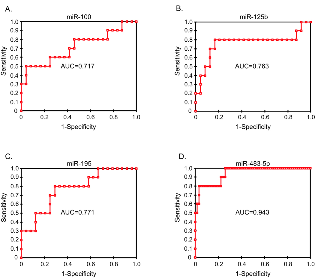
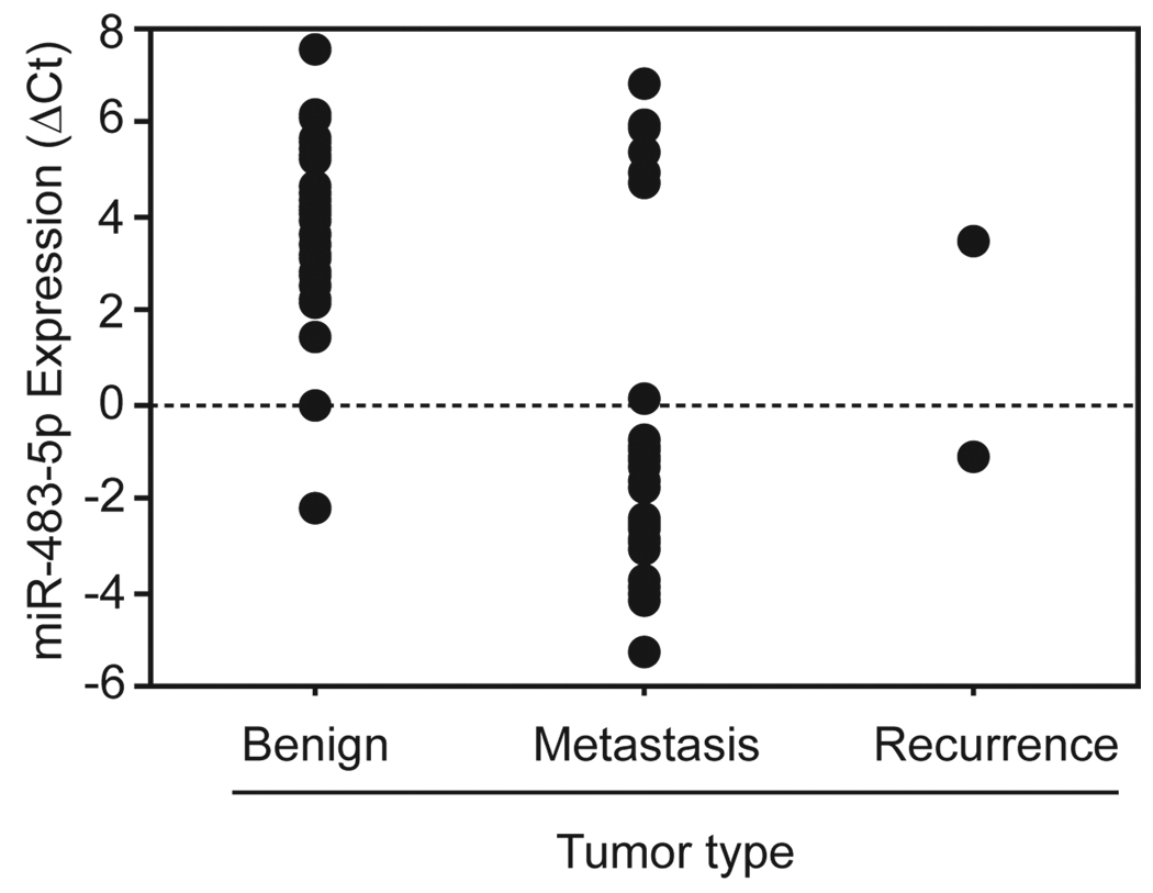
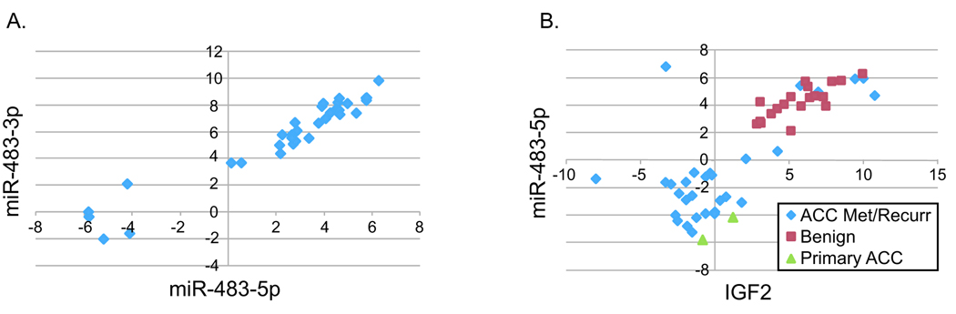
Results: Microarray profiling in benign and primary malignant adrenocortical tumors revealed several significant differences between these histological groups. By using directed quantitative RT-PCR analysis on a subset of these differentially expressed miRNAs, the authors determined that miRs -100, -125b, and -195 were significantly down-regulated, whereas miR-483-5p was significantly up-regulated in malignant as compared with benign tumors. Furthermore, the current study shows that miR-483-5p expression can accurately categorize tumors as benign or malignant.

Conclusions: The authors identified 4 miRNAs that are dysregulated in adrenocortical carcinoma. The high expression of one of these, miR-483-5p, appears to be a defining characteristic of adrenocortical malignancies, and can thus be used to accurately distinguish between benign and malignant adrenocortical tumors.


Conflict of interest statement

Conflict of Interest: There are no financial disclosures.

Figures

Figure 1
Figure 1. The miRNAs significantly up- or down-regulated in malignant and/or benign adrenocortical tumors as compared to normal adrenocortical tissue
Microarray analysis compared tumors (benign or malignant) to normal adrenocortical tissue. Differentially expressed genes were defined as those that had a p-value <0.01 (Ho: there is no difference between expression in tumor and normal). 17 miRNAs were misexpressed in both benign and malignant tumors (listed in the box, green and red indicate lower and higher expression in tumors, respectively). The underlined miRNAs were chosen to be validated by real-time quantitative RT-PCR.
Figure 2
Figure 2. Identification of differentially expressed miRNAs using microarray analysis
A. Unsupervised clustering was performed on the 50 most variable miRNAs. The most variable miRNAs were defined by the greatest absolute deviation from the mean across all samples. The Pearson correlation coefficient was used as the similarity metric in this analysis. The heatmaps show the clustering between clinical samples (columns) and the intensity of miRNA expression (log2 (tumor/normal), rows). The tumor diagnosis, either benign (B) or malignant (M), was indicated below. The microarrays included probes for mouse (mmu), human (hsa), and unannotated (miRPLUS) miRNAs (Exiqon LNA miRNA array v. 11.0). B. The top miRNAs significantly up- or down-regulated in ACC as compared to benign adrenocortical tumors were identified as those that were statistically significantly different (p<0.01, Ho: there is no difference between the expression in benign and malignant tumors) and had an expression difference of at least two-fold (up or down) between the two tumor classes were classified as the most differentially expressed. 23 miRNAs fit this criteria and the fold change for each is plotted (negative values indicate decreased expression in malignant and positive values indicate increased expression in malignant). The underlined miRNAs were chosen to be validated by real-time quantitative RT-PCR.
Figure 3
Figure 3. Real-time quantitative RT-PCR validation
Real-time quantitative RT-PCR was used to assay the expression of various miRNAs in tumor samples. The expression of each miRNA is expressed as the ΔCt (Ct miR of interest – Ct RNU48). The mean expression was calculated for benign tumors (n=24, except miRs-483-5p, 193b, and −125-5p where n=23) and ACCs (n=10) and is plotted (± standard error of the mean). An increase in ΔCt indicated lower expression whereas a decrease in ΔCt indicated higher expression (compare benign to malignant). A statistically significant difference between benign and malignant was indicated with an asterik (*) (p<0.05, Mann Whitney U test).
Figure 4
Figure 4. MiR-483-5p expression can distinguish benign from malignant tumors
The receiver operating characteristic curve (ROC) was plotted based on the real-time PCR expression profiling of miRs −100 (A), −125b (B), −195 (C), and −483-5p (D) (normalized to RNU48) for 34 samples (10 primary ACCs and 24 benign adrenocortical tumors). The area under the curve (AUC) was listed on the graph. With an AUC of 0.95, miR-483-5p had the greatest diagnostic accuracy (a perfect diagnostic marker without any false- negative or false-positives would have an AUC of 1).
Figure 5
Figure 5. High miR-483-5p expression is associated with adrenocortical malignancy
Real-time quantitative RT-PCR was used to assay the expression of miR-483-5p in tumor samples, either benign (n=35) or malignant (ACC recurrence, n=2; ACC metastasis, n=29). Each individual point on the plot denotes a patient sample. The y-axis represents the expression of miR-483-5p which was expressed as the ΔCt (Ct miR-483-5p – Ct RNU48). A lower ΔCt (compare benign to malignant) indicated higher miRNA expression. There was a statistically significant difference between the benign and malignant groups (p<0.0001, Mann Whitney U test).
Figure 6
Figure 6. The expression of miR-483-3p, miR-483-5p, and IGF2 mRNA are highly correlated
A. Real-time quantitative RT-PCR was used to assay the expression of miR-483-3p in tumor samples, either benign (n=24) or malignant (primary, n=10). MiR-483-3p expression, expressed as the ΔCt (Ct miR-483-3p – Ct RNU48), is plotted against the ΔCt of miR-483-5p for each patient sample. A strong positive correlation was detected (r=0.965, Pearson correlation). B. Real-time quantitative RT-PCR was used to assay the expression of IGF2 mRNA in tumor samples, either benign (n=19) or malignant (primary, n=2; ACC metastasis, n=27; ACC recurrence, n=3). IGF2 expression, as the ΔCt (Ct IGF2 – Ct GAPDH), is plotted against the ΔCt of miR-483-5p (Ct miR-483-5p – Ct RNU48) for each patient sample. A significant positive correlation was detected (r=0.799, Pearson correlation).

Similar articles

Cited by

References

    1. Icard P, Goudet P, Charpenay C, Andreassian B, Carnaille B, Chapuis Y, et al. Adrenocortical carcinomas: surgical trends and results of a 253-patient series from the French Association of Endocrine Surgeons study group. World J Surg. 2001. pp. 891–897. Available from http://www.ncbi.nlm.nih.gov/entrez/query.fcgi?cmd=Retrieve&db=PubMed&dop.... - PubMed
    1. Favia G, Lumachi F, Carraro P, D'Amico DF. Adrenocortical carcinoma. Our experience. Minerva Endocrinol. 1995. pp. 95–99. Available from http://www.ncbi.nlm.nih.gov/entrez/query.fcgi?cmd=Retrieve&db=PubMed&dop.... - PubMed
    1. Wajchenberg BL, Albergaria Pereira MA, Medonca BB, Latronico AC, Campos Carneiro P, Alves VA, et al. Adrenocortical carcinoma: clinical and laboratory observations. Cancer. 2000. pp. 711–736. Available from http://www.ncbi.nlm.nih.gov/entrez/query.fcgi?cmd=Retrieve&db=PubMed&dop.... - PubMed
    1. Brunt LM, Moley JF. Adrenal incidentaloma. World J Surg. 2001. pp. 905–913. Available from http://www.ncbi.nlm.nih.gov/entrez/query.fcgi?cmd=Retrieve&db=PubMed&dop.... - PubMed
    1. Lau SK, Weiss LM. The Weiss system for evaluating adrenocortical neoplasms: 25 years later. Hum Pathol. 2009. pp. 757–768. Available from http://www.ncbi.nlm.nih.gov/entrez/query.fcgi?cmd=Retrieve&db=PubMed&dop.... - PubMed

Publication types

MeSH terms