. 2011 Jul-Aug;24(4):585-96.
doi: 10.1002/jmr.1080. Epub 2010 Dec 13.

Binding of the cSH3 domain of Grb2 adaptor to two distinct RXXK motifs within Gab1 docker employs differential mechanisms

Affiliations

Binding of the cSH3 domain of Grb2 adaptor to two distinct RXXK motifs within Gab1 docker employs differential mechanisms

Caleb B McDonald et al. J Mol Recognit. 2011 Jul-Aug.

Abstract

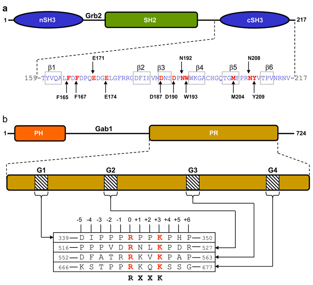
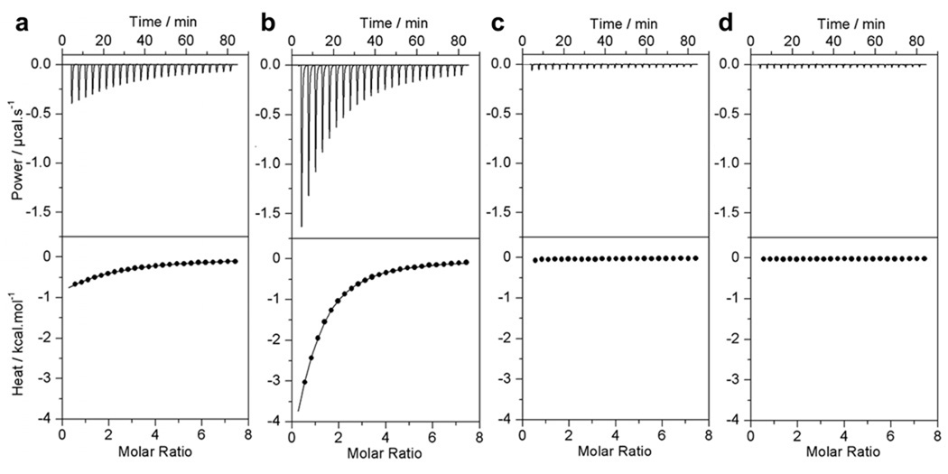
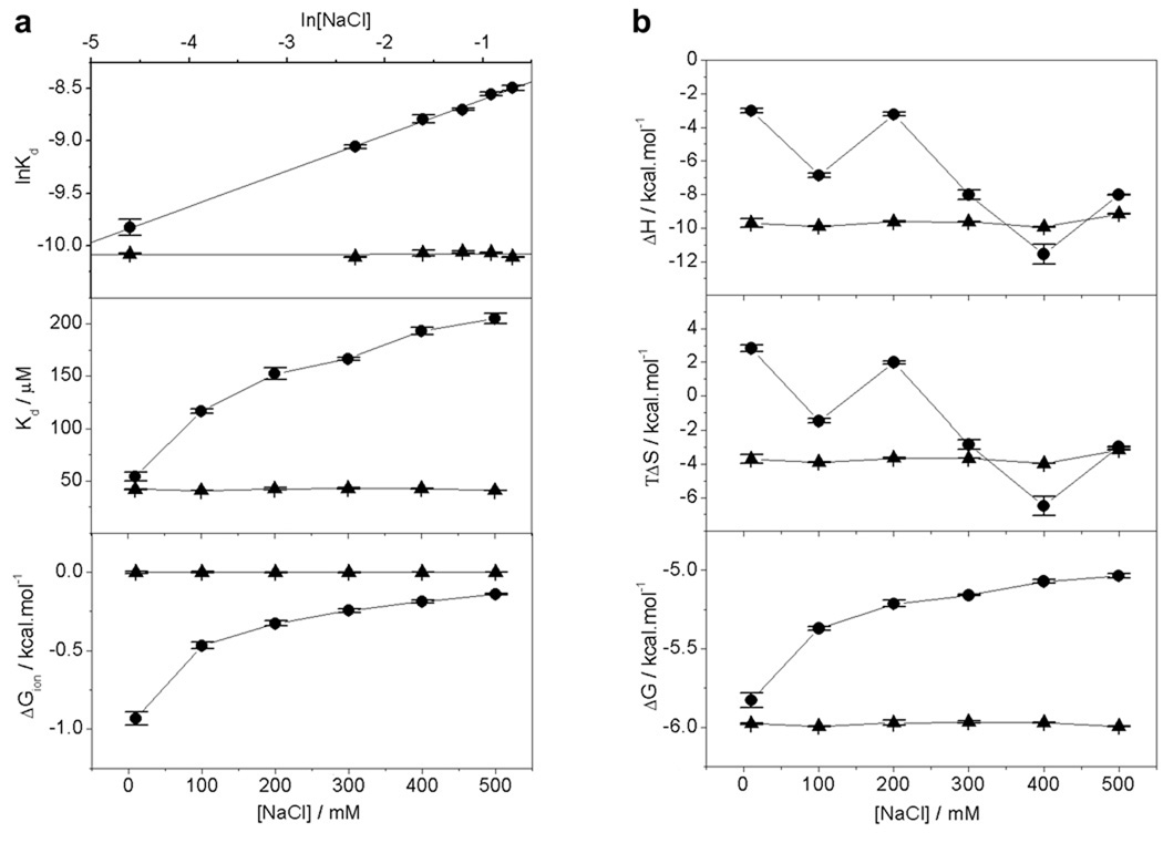
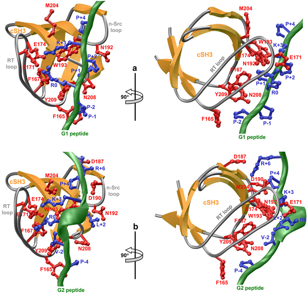
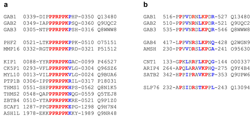
A ubiquitous component of cellular signaling machinery, Gab1 docker plays a pivotal role in routing extracellular information in the form of growth factors and cytokines to downstream targets such as transcription factors within the nucleus. Here, using isothermal titration calorimetry (ITC) in combination with macromolecular modeling (MM), we show that although Gab1 contains four distinct RXXK motifs, designated G1, G2, G3, and G4, only G1 and G2 motifs bind to the cSH3 domain of Grb2 adaptor and do so with distinct mechanisms. Thus, while the G1 motif strictly requires the PPRPPKP consensus sequence for high-affinity binding to the cSH3 domain, the G2 motif displays preference for the PXVXRXLKPXR consensus. Such sequential differences in the binding of G1 and G2 motifs arise from their ability to adopt distinct polyproline type II (PPII)- and 3(10) -helical conformations upon binding to the cSH3 domain, respectively. Collectively, our study provides detailed biophysical insights into a key protein-protein interaction involved in a diverse array of signaling cascades central to health and disease.


Figures

Figure 1
Figure 1
Modular organization of Grb2 adaptor and Gab1 docker. (a) Grb2 is comprised of a central SH2 (Src homology 2) domain flanked between an N-terminal SH3 (nSH3) domain and a C-terminal SH3 (cSH3) domain. The amino acid sequence of the cSH3 domain is indicated with the residues constituting the β1–β6 strands clearly demarcated. Key amino acid residues within the cSH3 domain involved in recognition of cognate ligands are colored red and labeled for clarity. (b) Gab1 is constructed on an N-terminal PH (Pleckstrin homology) domain and a C-terminal proline-rich (PR) domain separated by a long stretch of uncharacterized region. The PR domain contains four distinct RXXK motifs, here designated G1, G2, G3 and G4. The amino acid sequence of these motifs and flanking residues within Gab1 is provided. The numbering of various residues within and flanking the RXXK motifs is based on the nomenclature suggested by Feller and co-workers (Harkiolaki et al., 2009).
Figure 2
Figure 2
Representative ITC isotherms for the binding of cSH3 domain of Grb2 to Gab1-derived peptides G1 (a), G2 (b), G3 (c) and G4 (d). The upper panels show the raw ITC data expressed as change in thermal power with respect to time over the period of titration. In the lower panels, change in molar heat is expressed as a function of molar ratio of corresponding Gab1 peptide to cSH3 domain of Grb2. The solid lines in the lower panels show the fit of data to a one-site model, as embodied in Eq [1], using the ORIGIN software. Note that all data are shown to same scale for direct comparison.
Figure 3
Figure 3
Effect of salt on the energetics of binding of cSH3 domain of Grb2 to Gab1-derived peptides G1 and G2 as analyzed by ITC. (a) lnKd versus ln[NaCl] plots (upper panel), Kd versus [NaCl] plots (middle panel) and ΔGion versus [NaCl] plots (lower panel) for the binding of cSH3 domain to G1 peptide (●) and G2 peptide (▲). In the upper panel, the solid lines show linear fits to the data points, while solid lines in the middle and lower panels are merely used to connect data points for clarity. (b) Dependence of thermodynamic parameters ΔH, TΔS and ΔG on [NaCl] for the binding of cSH3 domain to G1 peptide (●) and G2 peptide (▲). The solid lines in all panels are merely used to connect data points for clarity. Each data point is the arithmetic mean of 3–4 independent experiments. All error bars are given to one standard deviation.
Figure 4
Figure 4
3D structural models of the cSH3 domain of Grb2 in complex with Gab1-derived peptides G1 and G2. (a) Ribbon representation of the cSH3 domain bound to G1 peptide. The β-strands in the cSH3 domain are shown in yellow with loops depicted in gray and the sidechains of key residues involved in making close contacts with the G1 peptide in red. The backbone of G1 peptide is colored green with the sidechains of all residues within the PPRPPKP motif depicted in blue. (b) Ribbon representation of the cSH3 domain bound to G2 peptide. The β-strands in the cSH3 domain are shown in yellow with loops depicted in gray and the sidechains of key residues involved in making close contacts with the G2 peptide in red. The backbone of G2 peptide is colored green with the sidechains of conserved residues within the PXVXRXLKPXR motif depicted in blue.
Figure 5
Figure 5
Amino acid sequence alignment showing the occurrence of PPRPPKP (a) and PXVXRXLKPXR (b) motifs in human proteome as identified through ScanProsite search at Expasy online server. Absolutely conserved residues within the motifs are shown in red, non-conserved residues within the motifs are depicted in blue, and all other residues are colored black. The proteins containing these motifs are listed in the left column and their corresponding Expasy codes are provided in the right column. The numerals hyphenated to amino acid sequence at each end denote the residue number within the protein sequence. Note that the amino acid sequences of PHF2 and MMP16 containing the PPRPPKP motif in (a) are provided in retro.

Similar articles

Cited by

References

    1. Alkarain A, Jordan R, Slingerland J. p27 deregulation in breast cancer: prognostic significance and implications for therapy. J Mammary Gland Biol Neoplasia. 2004;9:67–80. - PubMed
    1. Alkarain A, Slingerland J. Deregulation of p27 by oncogenic signaling and its prognostic significance in breast cancer. Breast Cancer Res. 2004;6:13–21. - PMC - PubMed
    1. Araki T, Nawa H, Neel BG. Tyrosyl phosphorylation of Shp2 is required for normal ERK activation in response to some, but not all, growth factors. J Biol Chem. 2003;278:41677–41684. - PubMed
    1. Barnett P, Bottger G, Klein AT, Tabak HF, Distel B. The peroxisomal membrane protein Pex13p shows a novel mode of SH3 interaction. Embo J. 2000;19:6382–6391. - PMC - PubMed
    1. Bennett HL, Brummer T, Jeanes A, Yap AS, Daly RJ. Gab2 and Src co-operate in human mammary epithelial cells to promote growth factor independence and disruption of acinar morphogenesis. Oncogene. 2008;27:2693–2704. - PubMed

Publication types

MeSH terms