. 2011 Apr 18;50(17):4003-6.
doi: 10.1002/anie.201008019. Epub 2011 Mar 29.

From crystal packing to molecular recognition: prediction and discovery of a binding site on the surface of polo-like kinase 1

Affiliations
Free PMC article

From crystal packing to molecular recognition: prediction and discovery of a binding site on the surface of polo-like kinase 1

Paweł Śledź et al. Angew Chem Int Ed Engl. .
Free PMC article
No abstract available


Figures

Figure 1
Figure 1
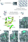
a) Upper: A simplified representation of the packing of PBD (gray)–peptide (purple) complexes in a crystal. Lower: Translation of the crystal-packing interaction into a two-pocket binder. b) Seven residues forming the new site in different conformations observed in the crystal structures; mobile residues shown in yellow. c) The closed hydrophobic pocket from our structure of the unligated PBD. d) The surface of the hydrophobic pocket from our structure of the PBD–MQSpSPL complex. Tyr481PBD and Tyr417PBD change the conformation to accommodate Leu394SYM; Leu can also be seen in the pocket in one of the previously published crystal structures. e) The surface of the hydrophobic pocket from our new structure of the PBD–MQSpTPL complex. Tyr481 is in an open conformation to accommodate Phe8term and Pro6term. f) The surface of the hydrophobic pocket from our structure of the PBD–PLHSpTA complex. Tyr417PBD and Phe482PBD change their conformations to accommodate Pro6term.
Figure 2
Figure 2
Comparison of the search string used to identify potential hydrophobic pocket-binding proteins and the matched sequences in PBIP1 and TCERG1. X, spacer residues; Φ, hydrophobic residue.
Figure 3
Figure 3
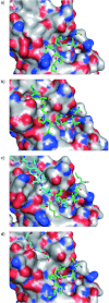
The structures of the PBD in complex with a) DPPLHSpTA; b) FDPPLHSpTA; c) MQSpTPL (the crystal contact with phenylalanine in the pocket was first observed in this structure—see Figure 1 e); d) FMPPPMSpSM peptides. The peptides are shown in green and the surface of the PBD is shown, flexible N-terminal tail of symmetry-related protomer in c) and polyethylene glycol in d) are shown in cyan.

References

    1. Wells JA, McClendon CL. Nature. 2007;450:1001–1009. - PubMed
    1. Whitty A, Kumaravel G. Nat. Chem. Biol. 2006;2:112–118. - PubMed
    1. DeLano WL, Ultsch MH, de Vos AM, Wells JA. Science. 2000;287:1279–1283. - PubMed
    1. Atwell S, Ultsch M, de Vos AM, Wells JA. Science. 1997;278:1125–1128. - PubMed
    1. Keskin O, Gursoy A, Ma B, Nussinov R. Chem. Rev. 2008;108:1225–1244. - PubMed

Publication types

LinkOut - more resources