. 2011 Jun 10;317(10):1368-81.
doi: 10.1016/j.yexcr.2011.03.019. Epub 2011 Apr 5.

The Notch ligand Delta-like 1 integrates inputs from TGFbeta/Activin and Wnt pathways

Affiliations

The Notch ligand Delta-like 1 integrates inputs from TGFbeta/Activin and Wnt pathways

Michael Bordonaro et al. Exp Cell Res. .

Abstract

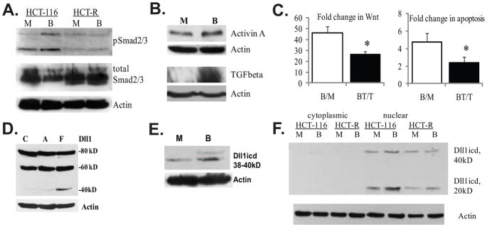
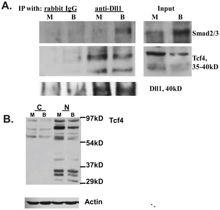
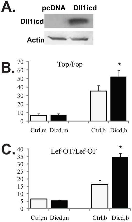
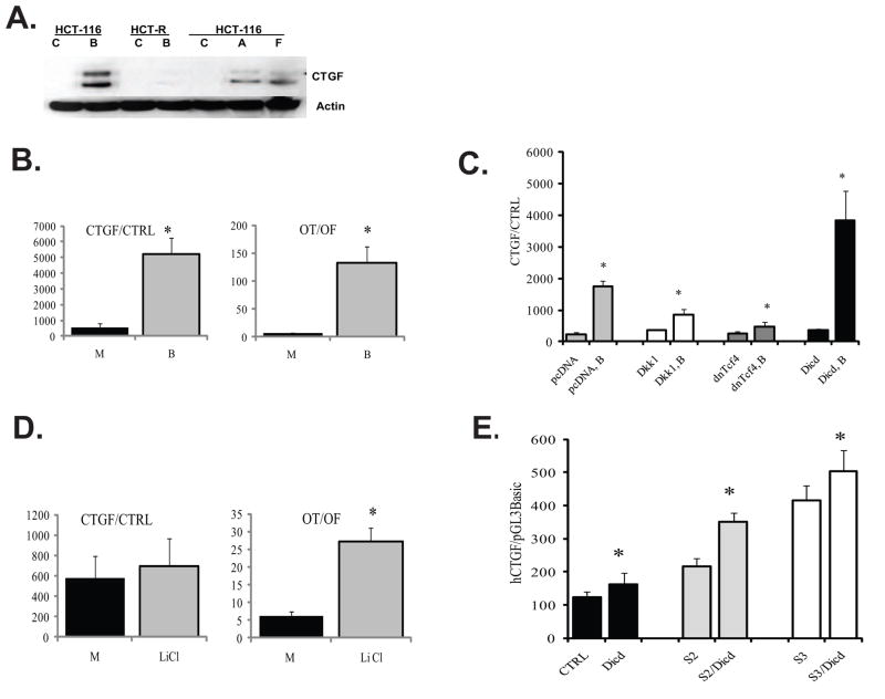
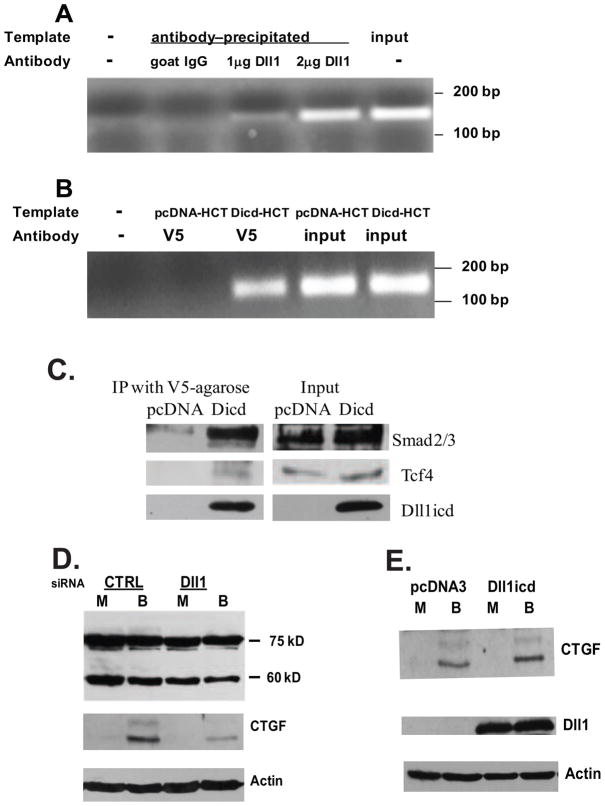
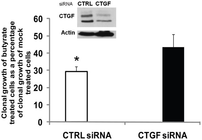
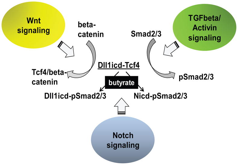
Unlike the well-characterized nuclear function of the Notch intracellular domain, it has been difficult to identify a nuclear role for the ligands of Notch. Here we provide evidence for the nuclear function of the Notch ligand Delta-like 1 in colon cancer (CC) cells exposed to butyrate. We demonstrate that the intracellular domain of Delta-like 1 (Dll1icd) augments the activity of Wnt signaling-dependent reporters and that of the promoter of the connective tissue growth factor (CTGF) gene. Data suggest that Dll1icd upregulates CTGF promoter activity through both direct and indirect mechanisms. The direct mechanism is supported by co-immunoprecipitation of endogenous Smad2/3 proteins and Dll1 and by chromatin immunoprecipitation analyses that revealed the occupancy of Dll1icd on CTGF promoter sequences containing a Smad binding element. The indirect upregulation of CTGF expression by Dll1 is likely due to the ability of Dll1icd to increase Wnt signaling, a pathway that targets CTGF. CTGF expression is induced in butyrate-treated CC cells and results from clonal growth assays support a role for CTGF in the cell growth-suppressive role of butyrate. In conclusion, integration of the Notch, Wnt, and TGFbeta/Activin signaling pathways is in part mediated by the interactions of Dll1 with Smad2/3 and Tcf4.


Figures

Fig. 1
Fig. 1
Evidence for TGFbeta/Activin signaling and expression of Dll1 species in HCT-116 colon cancer cells. (A). Butyrate induces the phosphorylation of Smad2/3 proteins in butyrate-sensitive HCT-116, but not in butyrate-resistant HCT-R cells. A representative Western blot of total and phosphorylated Smad2/3 in HCT-116 and HCT-R cells exposed to 24h of mock (M) or 5 mM sodium butyrate (B) treatment. The anti-phospho Smad2/3 antibody (sc-11769-R, Santa Cruz Biotechnology) recognizes the phosphorylated forms of both Smad2 and Smad3 (53kD and 60kD). (B). TGFbeta and Activin A expression is augmented in butyrate-treated HCT-116 cells. A representative Western blot analysis of Activin A expression in HCT-116 cells exposed to a 24-h mock (M) or 5 mM of sodium butyrate (B) treatment. Goat anti-Activin A antibody (R&D Systems, #AF338, used at 0.4 μg/ml), recognizes the Activin A dimer (28 kD), consisting of two inhibin beta-chains, as described by others [37]. (C) Suppression of TGFbeta/Activin signaling counteracts the induction of Wnt activity and apoptosis in butyrate-treated HCT-116 cells. Cells were transfected with the Wnt-sensitive luciferase reporters Lef-OT or Lef-OF, and the pRL-null vector, added as a measure for transfection efficiency. Transfected cells were exposed for 24h to mock treatment (M), 5mM butyrate (B), 20μM TGFbeta/Activin inhibitor SB-505124 from Santa Cruz Biotechnology (T), or 20μM inhibitor and 5mM butyrate (BT). Luciferase activity was assayed with dual luciferase kit (Promega), and the ratios of LefOT/LefOF, indicative of Wnt transcriptional activity were calculated. The B/M ratio is the fold increase of Wnt activity in butyrate- vs. mock-treated cells (625.0±72.7/13.9±3.49); whereas the BT/T ratio is the fold increase of Wnt activity in butyrate+inhibitor-treated cells versus inhibitor-treated cells (399.4±79.2/15.9±4.56), P<0.05. The data are the mean of three independent experiments with duplicate samples in each experiment. Apoptosis was measured in cells exposed to the same treatments (see Materials and Methods). The ratio B/M is the ratio of the percentages apoptotic cells in butyrate- vs. mock-treated samples (4.74±1.02); the ratio BT/T is the ratio of the percentages apoptotic cells in butyrate+inhibitor- treated samples vs. inhibitor-treated samples 2.41±0.62, P<0.05. We analyzed three samples per treatment in each of three independent experiments. Bars, SDs. (D). Highest levels of a Dll1icd-containing species are detected in floating fractions of butyrate-treated HCT-116 cells. Exposure of HCT-116 cells to 5 mM sodium butyrate for 48h results in a fraction of floating cells (F) and a fraction of substrate-adherent cells (A). Total protein lysates (150 μg) from these two cell fractions and mock-treated cells (C) were analyzed by Western blotting. The anti-Dll1 antibody detects the full length ligand (80kD), a form of the ligand with partially shed ectodomain (60kD), and a carboxy-terminal form of 38–40kD. (E) Butyrate-treated HCT-116 cells express higher levels of carboxy-terminal 38–40kD Dll1 species compared to mock-treated cells. A representative Western blot analysis of nuclear lysates (80 μg) of HCT-116 cells exposed to mock or 5 mM sodium butyrate treatment for 17h. Detection was achieved with a goat anti-Dll1 antibody (sc-8155, Santa Cruz Biotechnology) and a beta-Actin antibody (Sigma). (F) Highest levels of Dll1icd (20kD) are detected in nuclear fractions of butyrate-treated HCT-116 cells. Cytoplasmic-nuclear fractionation of HCT-116 cells exposed to mock (M) or 5 mM butyrate (B) treatment for 17h was performed with the Pierce Nuclear and Cytoplasmic Extraction Reagent Kit (NE-PER). A total of 100 μg of cytoplasmic or nuclear proteins were analyzed on 12% SDS gels. Western blot detection of Dll1 species was carried out with a rabbit anti-Dll1 antibody (sc-9102, Santa Cruz Biotechnology).
Fig. 2
Fig. 2
Dll1 associates with Smad2/3 and Tcf4 in lysates of HCT-116 CC cells. (A) Immunoprecipitation (IP) analyses with an anti-Dll1 antibody. Nuclear protein lysates (100 μg) from HCT-116 cells, exposed to mock (M) or 5 mM butyrate (B) treatment for 17h, were used for IPs with 2 μg of normal IgG (control) or with 2μg of anti-Dll1 antibody (sc-9102, Santa Cruz Biotechnology). Smad2/3 proteins were detected with a polyclonal antibody that recognizes both proteins (sc-8332, Santa Cruz Biotechnology), Tcf4 was detected with a monoclonal antibody from Millipore (clone 6H5-3). Control experiments confirmed that the anti-Dll1antibody immunoprecipitates the 40kD form of Dll1; we were unable to detect this form in the input, due to its low abundance. (B) Nuclear (N), but not cytoplasmic (C) fractions of HCT-116 cells exhibit the presence of low molecular weight Tcf4 isoforms (30–45 kD). Fractionation of HCT-116 cells exposed to mock (M) or 5 mM butyrate (B) treatment for 24h was performed with the Pierce Nuclear and Cytoplasmic Extraction Reagent Kit (NE-PER). Detection of Tcf4 was carried out as in (A).
Fig. 3
Fig. 3
The butyrate-induced Wnt activity is augmented by Dll1icd in HCT-116 CC cells. (A) Representative Western blot analyses of HCT-116 CC cells transiently transfected with pcDNA3.1 or a construct expressing Dll1icd. The exogenous protein was detected with a Dll1-specific antibody (sc-8155, Santa Cruz Biotechnology). (B) Dll1icd augments the induction of Wnt signaling by butyrate, as measured with the TopFlash and FopFlash reporter constructs. HCT-116 cells were co-transfected transiently with one of the Wnt signaling reporters (TopFlash or FopFlash), pcDNA3.1 or Dll1icd-expression vector in a 96-well plate. pRL-TK was co-transfected as a measure for transfection efficiency. The ratio between the reporters and the effector vectors was 1:3, the total amount DNA per well was 0.32 μg. Transfections were carried out with Lipofectamine 2000, and treatment with 5 mM butyrate was for 17h. Each transfection was performed in duplicate wells; data represent the mean from results of at least three experiments. Wnt signaling was calculated as the ratio TopFlash/FopFlash (Top/Fop). There was no statistically significant difference between Wnt activity in mock-treated cells transfected with pcDNA3.1 or Dll1icd-expressing construct. For butyrate-treated cells, Top/Fop was 35.3±6.6 and 52.4±6.9 (P<0.05) for pcDNA- and Dll1icd-transfected cells, respectively. (C) Dll1icd augments the induction of Wnt signaling by butyrate, as measured with the Lef-OT and Lef-OF reporter constructs. The transfections were performed as described in 3B. The ratio Lef-OT/Lef-OF is a measure of Wnt activity. There was no significant difference between Wnt levels in mock-treated cells transfected with empty vector or Dll1icd-expressing construct. In butyrate-treated cells, Lef-OT/Lef-OF was 16.3±2.7 and 34.8±2.4 (P<0.05) for pcDNA- and Dll1icd-transfected cells, respectively.
Fig. 4
Fig. 4
Butyrate induces CTGF expression in HCT-116 cells, and this effect is modulated by components of Wnt signaling and Dll1icd. (A) CTGF expression is induced by butyrate in butyrate-sensitive HCT-116 cells, but not in butyrate-resistant HCT-R cells. HCT-116 and HCT-R cells were exposed to mock (C) or 5 mM butyrate (B) treatment for 24h. In addition, HCT-116 cells were treated for 48h with 5 mM butyrate, and lysates from the floating (F) and substrate-adherent (A) cells were analyzed along with lysates of mock-treated (C) cells. Total protein lysates (100μg) were analyzed by Western blot analyses, CTGF and beta-Actin were detected with antibodies from Santa Cruz Biotechnology (sc-14939) and Sigma (A5441), respectively. The production of two CTGF isoforms of 35 and 38 kD, and smaller proteolytically processed forms have been previously reported [48]. (B) Butyrate treatment of HCT-116 cells upregulates the activity of the CTGF promoter. HCT-116 cells were co-transfected with the CTGF reporter and the normalization plasmid pRL-TK, or with the promoterless pGL3Basic and pRL-TK, in a 96-well plate format, following the Lipofectamine 2000 quick transfection protocol (see Materials and Methods). Cells were assayed for luciferase activity after 17-h exposure to mock (M) or 5 mM butyrate (B) treatment. For comparative analyses, the same transfection protocol was applied for Wnt signaling reporters (Lef-OT and Lef-OF). Each transfection was performed in duplicate wells; data represent the mean from results of at least three experiments. (C) Exogenous expression of Dll1icd, dnTcf4, and Dkk1 modulate the induction of CTGF by butyrate. HCT-116 cells were co-transfected with the CTGF promoter reporter (CTGF) or with its promoterless version (CTRL) and one of the following plasmids: pcDNA3Neo (empty vector), Dkk1-expression vector, dnTcf4-expression vector, or Dll1icd (Dicd)-expression vector. All transfections included pRL-TK as a control for transfection efficiency. We applied the quick transfection protocol for Lipofectamine 2000 in 96-well plates. Approximately 60,000 cells were transfected with reporter to effector plasmid ratio at 1:3, to a total of 320ng DNA per well and 0.8 μl of Lipofectamine. After six hours of incubation with the transfection mixture, cells were exposed to mock or 5 mM butyrate treatment for 17h. The ratio of CTGF promoter activity to that of the promoterless reporter was calculated for cells in absence or presence of butyrate. Data represent the mean from results of at least three transfections; each transfection was performed in duplicate wells. There were statistically significant differences between the CTGF promoter activities in butyrate-treated cells expressing Dkk1, dnTcf4, or Dicd and the CTGF promoter activity in butyrate-treated cells transfected with an empty pcDNA vector, (P-values were 0.001, 0, and 0.021, respectively). (D) Increase in active (dephosphorylated) beta-catenin is not sufficient for the induction of CTGF by butyrate. HCT-116 cells were transfected with CTGF or Wnt reporters as described in (B) and exposed to 20mM lithium chloride (LiCl) for 17h. Each transfection was performed in duplicate wells; data represent the mean from results of at least three experiments. There was no statistically significant difference between the activities of the CTGF promoter in mock- and LiCl-treated cells (P>0.05); however, control transfection experiments confirmed the ability of LiCl to induce the Wnt-sensitive promoter Lef-OT (P<0.05). (E) Co-expression of Dll1icd (Dicd) with constitutively active (ca) Smad2 (S2) or constitutively active Smad3 (S3) activates the CTGF promoter in the absence of butyrate. HCT-116 cells were co-transfected with the CTGF promoter reporter (CTGF) or the control pGL3Basic, and one of the following combinations of plasmids: pcDNA3Neo (empty vector), Dll1icd (Dicd)-expression vector, Dll1icd and caSmad2, or Dll1icd and caSmad3. We utilized the reverse Lipofectamine protocol in a 96-well format; the reporters were used at 80 ng per well, the effectors were used at total of 240 ng. pRL-TK was co-transfected at 1:200 ratio for transfection efficiency. Fresh medium was added at five hours post-transfection, and luciferase assays were performed at 24h post-transfection. Data represent the mean from results of at least three transfections; each transfection was performed in duplicate wells. Bars are SDs; statistically significant differences are marked with asterisks.
Fig. 5
Fig. 5
Dll1icd modulates directly the activity of the CTGF promoter in butyrate-treated HCT-116 cells. (A) Endogenous Dll1 protein species is associated with the CTGF promoter region. Chromatin immunoprecipitation (ChIP) analyses were performed with goat anti-Dll1 or normal goat IgG preparations (Santa Cruz Biotechnology) and lysates of butyrate-treated HCT-116 cells, as described in Materials and Methods. One-fifth of each immunoprecipitation reaction was used as a template for the PCR with CTGF promoter-specific primers. One-half of the PCR products were analyzed on 1.7% agarose gels. On panel A, lane 1 has no DNA template, lane 2 - template precipitated with 2 μg of normal goat IgG, lane 3 - template precipitated with 1 μg of anti-Dll1 antibody, lane 4 - template precipitated with 2 μg of anti-Dll1 antibody, lane 5 – input (0.2% of the input for the ChIP reactions). The PCR product is 143 nucleotides long. (B) Exogenously expressed Dll1icd is associated with the CTGF promoter. ChIP analyses were performed with anti-V5 beads (Sigma) and lysates of butyrate-treated HCT-116 cells nucleofected with an empty vector (pcDNA, control) or with a Dll1icd-expression construct. On panel B, lane 1 has no DNA template, lane 2 - template precipitated from pcDNA-transfected HCT-116 cells, lane 3 - template precipitated from Dicd-transfected HCT-116 cells, lane 4 - template from input, pcDNA-transfected cells, lane 5 - template from input, Dll1icd-transfected cells. (C) Exogenous Dll1icd associates with Smad2/3 and Tcf4 proteins in ChIP lysates of butyrate-treated HCT-116 cells. ChIP lysates from cells transfected with empty vector (pcDNANeo) or Dll1icd-expressing constract were immunoprecipitated with V5-agarose beads (Sigma). The precipitated material was eluted from the beads by adding 2x Laemmli buffer and analyzed via Western blotting. The antibodies for total Smad2/3 and Tcf4 are described in the legend of Fig.2A. (D) Downregulation of endogenous Dll1 levels results in a suppressed induction of CTGF. Nucleofection of HCT-116 cells was carried out as described in Materials and Methods. Cells were exposed to mock treatment or 5 mM butyrate for 17h and total cell lysates were analyzed by Western blotting with a CTGF antibody. (E). Overexpression of Dll1icd in HCT-116 cells results in enhanced induction of CTGF. Dll1icd was overexpressed in HCT-116 cells were nucleofected with pcDNA or Dll1icd expression vector, exposed to mock or 5 mM butyrate for 17h, and total cell lysates were analyzed via Western blotting for CTGF protein expression. The signal for beta-Actin was used as a loading control.
Fig. 6
Fig. 6
Suppressed CTGF expression contributes to the resistance of HCT-116 cells to the growth inhibitory effects of butyrate. HCT-116 cells (106) were nucleofected with 175 pmol of control (non-specific) or human CTGF siRNA (Santa Cruz Biotechnology) and incubated in two wells of a 6-well plate. At six hours post-transfection the cells were mock-treated or exposed to 5 mM sodium butyrate for 17 hours. The inset is a representative Western blot analysis of the CTGF protein levels in control and CTGF siRNA-nucleofected cells exposed to 5 mM butyrate for 17h. Clonal growth assays were performed as described in Materials and Methods. The clonal growth of butyrate-treated cells is expressed as a percentage of the clonal growth of the mock-treated cells. The clonal growth assays were repeated at least three to six times. Statistically significant differences are marked with asterisks. Bars, SDs.
Fig. 7
Fig. 7
Integration of three signaling pathways. Wnt signaling results in formation of Tcf-4/beta- - catenin complexes which activate expression from Wnt targeted genes; TCFbeta/Activin signaling is mediated by phosphorylated Smad2/3. In mock-treated HCT-116 cells, a limited amount of Dll1icd is bound to Tcf4 species, and likely to dephosphorylated Smad2/3; these transcriptional complexes might be repressive. In butyrate-treated HCT-116 cells, the steady-state levels of the Tcf4 species decrease, and some of the Tcf4 protein pool is recruited to Wnt signaling complexes with dephosphorylated beta-catenin. Butyrate-treated cells also exhibit increased levels of phosphorylated Smad2/3 (pSmad2/3) proteins. In a fraction of the cell population, the pSmad2/3 proteins are bound to Dll1icd, and the resulting complexes may activate a specific set of genes (e.g., CTGF, p21, etc). In the remainder of the cell population, the pSmad2/3 proteins bind to Nicd, and these complexes may modulate a different set of genes [–82]. Our preliminary data suggest that cells with higher levels of Dll1icd respond to butyrate with greater hyper-activation of Wnt signaling, and higher levels of apoptosis. The cells with higher levels of Nicd may suppress the hyper-activation of Wnt/beta-catenin by butyrate, and are likely more resistant to apoptosis.

References

    1. Sancho E, Batlle E, Clevers H. Signaling pathways in intestinal development and cancer. Annu Rev Cell Dev Biol. 2004;20:695–723. - PubMed
    1. Morin J, Sparks AB, Korinek V, Barker N, Clevers H, Vogelstein B, Kinzler KW. Activation of beta-catenin-Tcf signaling in colon cancer by mutations in beta-catenin or APC. Science. 1997;275:1787–1790. - PubMed
    1. Kinzler KW, Vogelstein B. Lessons from hereditary colorectal cancer. Cell Biochem Biophys. 1996;87:159–170. - PubMed
    1. Miyaki M, Iijima T, Kimura J, Yasuno M, Mori T, Hayashi Y, Koike M, Shitara N, Iwama T, Kuroki T. Frequent mutation of beta-catenin and APC genes in primary colorectal tumors from patients with hereditary nonpolyposis colorectal cancer. Cancer Res. 1999;59:4506–4509. - PubMed
    1. Roose J, Clevers H. Tcf transcription factors: molecular switches in carcinogenesis. Biochim Biophys Acta. 1999;87456:M23–M27. - PubMed

Publication types

MeSH terms