

Extranuclear signaling of mutated thyroid hormone receptors in promoting metastatic spread in thyroid carcinogenesis
- PMID: 21473875
- PMCID: PMC3129395
- DOI: 10.1016/j.steroids.2011.03.016
Extranuclear signaling of mutated thyroid hormone receptors in promoting metastatic spread in thyroid carcinogenesis
Abstract
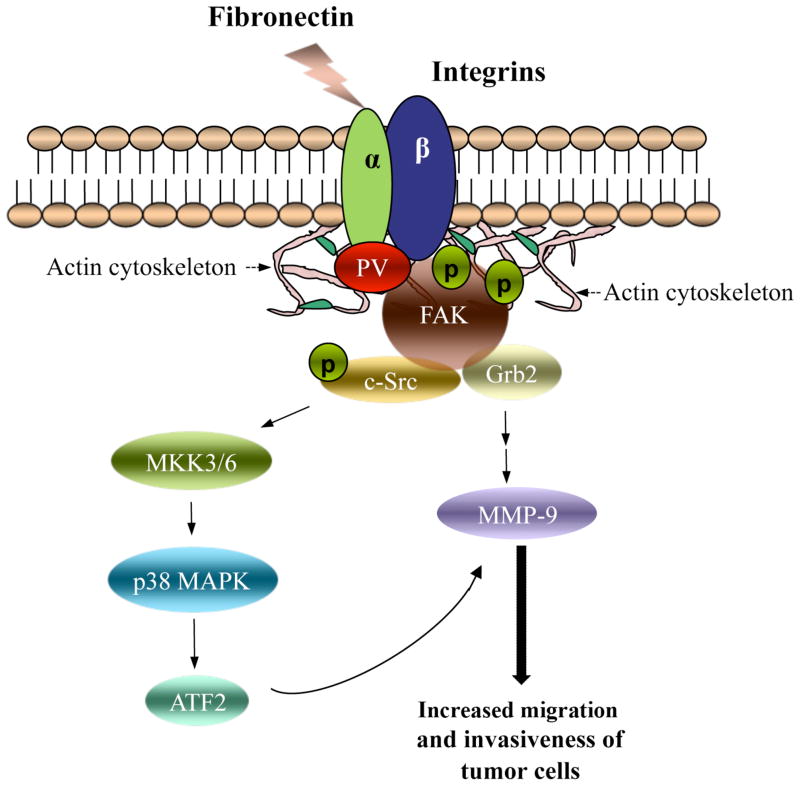
Thyroid hormone receptors (TRs) mediate the critical activities of the thyroid hormone (T3) in growth, development, and differentiation. Decreased expression and/or somatic mutations of TRs have been shown to be associated with several types of human cancers including liver, breast, lung, and thyroid. A direct demonstration that TRβ mutants could function as oncogenes is evidenced by the spontaneous development of follicular thyroid carcinoma similar to human cancer in a knockin mouse model harboring a mutated TRβ (denoted as PV; Thrb(PV/PV) mice). PV is a dominant negative mutation identified in a patient with resistance to thyroid hormone. Analysis of altered gene expression and molecular studies of thyroid carcinogenesis in Thrb(PV/PV) mice show that the oncogenic activity of PV is mediated by both nucleus-initiated transcription and extranuclear actions to alter gene expression and signaling transduction activity. This article focuses on recent findings of novel extranuclear actions of PV that affect signaling cascades and thereby the invasiveness, migration, and motility of thyroid tumor cells. These findings have led to identification of potential molecular targets for treatment of metastatic thyroid cancer.
Published by Elsevier Inc.
Figures
Similar articles
-
Inhibition of phosphatidylinositol 3-kinase delays tumor progression and blocks metastatic spread in a mouse model of thyroid cancer.Carcinogenesis. 2007 Dec;28(12):2451-8. doi: 10.1093/carcin/bgm174. Epub 2007 Jul 28. Carcinogenesis. 2007. PMID: 17660507
-
Activation of tumor cell proliferation by thyroid hormone in a mouse model of follicular thyroid carcinoma.Oncogene. 2012 Apr 19;31(16):2007-16. doi: 10.1038/onc.2011.390. Epub 2011 Sep 12. Oncogene. 2012. PMID: 21909131 Free PMC article.
-
Novel oncogenic actions of TRbeta mutants in tumorigenesis.IUBMB Life. 2009 May;61(5):528-36. doi: 10.1002/iub.180. IUBMB Life. 2009. PMID: 19391168 Free PMC article.
-
Nongenomic activation of phosphatidylinositol 3-kinase signaling by thyroid hormone receptors.Steroids. 2009 Jul;74(7):628-34. doi: 10.1016/j.steroids.2008.10.009. Epub 2008 Oct 30. Steroids. 2009. PMID: 19014961 Free PMC article. Review.
-
Novel non-genomic signaling of thyroid hormone receptors in thyroid carcinogenesis.Mol Cell Endocrinol. 2009 Sep 24;308(1-2):63-9. doi: 10.1016/j.mce.2009.01.007. Epub 2009 Jan 21. Mol Cell Endocrinol. 2009. PMID: 19549593 Free PMC article. Review.
Cited by
-
Bioactivity of Thyroid Hormone Analogs at Cancer Cells.Front Endocrinol (Lausanne). 2018 Dec 4;9:739. doi: 10.3389/fendo.2018.00739. eCollection 2018. Front Endocrinol (Lausanne). 2018. PMID: 30564196 Free PMC article. Review.
-
Role of TSH in the spontaneous development of asymmetrical thyroid carcinoma in mice with a targeted mutation in a single allele of the thyroid hormone-β receptor.Endocrinology. 2012 Oct;153(10):5090-100. doi: 10.1210/en.2012-1600. Epub 2012 Aug 23. Endocrinology. 2012. PMID: 22919057 Free PMC article.
-
Mechanisms of thyroid hormone action.J Clin Invest. 2012 Sep;122(9):3035-43. doi: 10.1172/JCI60047. Epub 2012 Sep 4. J Clin Invest. 2012. PMID: 22945636 Free PMC article. Review.
-
Thyroid hormone receptors and cancer.Biochim Biophys Acta. 2013 Jul;1830(7):3928-36. doi: 10.1016/j.bbagen.2012.04.002. Epub 2012 Apr 6. Biochim Biophys Acta. 2013. PMID: 22507269 Free PMC article. Review.
-
Small-molecule hormones: molecular mechanisms of action.Int J Endocrinol. 2013;2013:601246. doi: 10.1155/2013/601246. Epub 2013 Feb 28. Int J Endocrinol. 2013. PMID: 23533406 Free PMC article.
References
-
- Cheng SY. Multiple mechanisms for regulation of the transcriptional activity of thyroid hormone receptors. Rev Endocr Metab Disord. 2000;1:9–18. - PubMed
-
- Bassett JH, Harvey CB, Williams GR. Mechanisms of thyroid hormone receptor-specific nuclear and extra nuclear actions. Mol Cell Endocrinol. 2003;213:1–11. - PubMed
-
- O’Shea PJ, Williams GR. Insight into the physiological actions of thyroid hormone receptors from genetically modified mice. J Endocrinol. 2002;175:553–70. - PubMed
-
- Lin KH, Shieh HY, Chen SL, Hsu HC. Expression of mutant thyroid hormone nuclear receptors in human hepatocellular carcinoma cells. Mol Carcinog. 1999;26:53–61. - PubMed
Publication types
MeSH terms
Substances
Grants and funding
LinkOut - more resources
Full Text Sources
Other Literature Sources
Medical
