. 2011 Apr 8;88(4):508-15.
doi: 10.1016/j.ajhg.2011.03.015.

Human and mouse mutations in WDR35 cause short-rib polydactyly syndromes due to abnormal ciliogenesis

Affiliations

Human and mouse mutations in WDR35 cause short-rib polydactyly syndromes due to abnormal ciliogenesis

Pleasantine Mill et al. Am J Hum Genet. .

Abstract

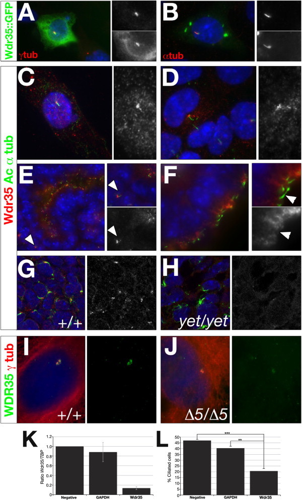
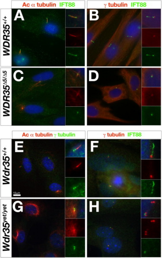
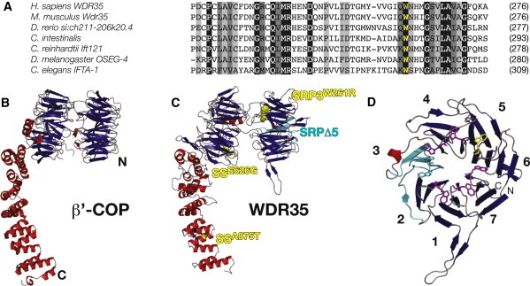
Defects in cilia formation and function result in a range of human skeletal and visceral abnormalities. Mutations in several genes have been identified to cause a proportion of these disorders, some of which display genetic (locus) heterogeneity. Mouse models are valuable for dissecting the function of these genes, as well as for more detailed analysis of the underlying developmental defects. The short-rib polydactyly (SRP) group of disorders are among the most severe human phenotypes caused by cilia dysfunction. We mapped the disease locus from two siblings affected by a severe form of SRP to 2p24, where we identified an in-frame homozygous deletion of exon 5 in WDR35. We subsequently found compound heterozygous missense and nonsense mutations in WDR35 in an independent second case with a similar, severe SRP phenotype. In a mouse mutation screen for developmental phenotypes, we identified a mutation in Wdr35 as the cause of midgestation lethality, with abnormalities characteristic of defects in the Hedgehog signaling pathway. We show that endogenous WDR35 localizes to cilia and centrosomes throughout the developing embryo and that human and mouse fibroblasts lacking the protein fail to produce cilia. Through structural modeling, we show that WDR35 has strong homology to the COPI coatamers involved in vesicular trafficking and that human SRP mutations affect key structural elements in WDR35. Our report expands, and sheds new light on, the pathogenesis of the SRP spectrum of ciliopathies.


Figures

Figure 1
Figure 1
WDR35 Is Mutated in Atypical Short-Rib Polydactyly Syndrome in Humans and in the yeti Mutant Mouse (A and B) Characteristic postaxial polydactyly, extreme micromelia, and short ribs (A) as presented in postmortem survey of 13 week conceptus (SRP-3-1) (B). (C) Genomic PCR spanning exons 4, 5, or 6 of WDR35 from DNA of parents (SRP-1-1, SRP-1-2) or affected concepti (SRP-1-4, SRP-1-6). Deletion of a 2847 bp genomic fragment (2:20177392-20180238, assembly GRCh37) results in the loss of exon 5 in homozygous individuals. Reaction products for the control (C1) and no template (-ve) are shown. (D) RT-PCR amplification of WDR35 spanning exons 4–6 from affected concepti (SRP-1-4, SRP-1-6) and control fibroblasts (C1, C2). (E) Schematic of the predicted domain organization of two coding human WDR35 transcripts, with or without exon 11 (violet). The location of the WDR35 mutations in SRP-1 (blue; homozygous SRPΔ5) and SRP-3 (p.Trp261Arg and p.Arg545X) -affected concepti are shown. The splice acceptor mutation at the intron 21-exon 22 junction in yeti mice is indicated (). (F–K) Gross embryonic phenotypes of wild-type (F–H) compared to yeti mutant littermates (I–K). Embryonic day 12.5 (E12.5) forelimb (F, I) and hindlimb (G, J) defects in yeti mouse mutants include impaired outgrowth along the proximal-distal axis and polysyndactyly. (H, K) E11.5 transverse hematoxylin- and eosin-stained sections of the Wdr35yet/yet thoracic cavity show failure of the somite derivatives, such as the sclerotome (sc) derivatives (ri, ribs; ve, vertebra), to migrate out from the midline in mutants and tracheoesophageal fistula with hypoplastic lungs (asterisk). (L) RT-PCR amplification of a Wdr35 fragment covering exons 21–24 shows the presence of aberrantly spliced of transcripts in the yeti mutant (white arrowheads) compared to wild-type (black arrowhead). (M) Whole-mount in situ hybridization of Wdr35 in E10.5 wild-type and yeti littermates shows nonsense-mediated decay of mRNA in mutant embryos.
Figure 2
Figure 2
Wdr35 Localizes to Cilia and Is Required for Ciliogenesis (A and B) NIH-3T3 (A) or IMCD3 (B) cells were microporated with full-length mouse Wdr35::GFP and serum starved for 36 hr before costaining with antibodies directed against γ-tubulin (A) or acetylated α-tubulin (red; B). Nuclei are stained with DAPI (blue). Magnification of regions of interest are shown in singl-channel images indicating colocalization. (C and D) IMCD3 cells were serum starved for 36 hr before costaining with antibodies directed against acetylated α-tubulin (green) and Wdr35 (red; C, 085 antibody; D, 02 antibody). Nuclei are stained with DAPI (blue). (E and F) Wild-type 13.5 dpc mouse kidney (E) and choroid plexus (F) sections were costained with antibodies directed against acetylated α-tubulin (green) and Wdr35 (red, 085 antibody). Nuclei are stained with TOTO-3 (blue). Primary cilia in the mesenchyme (E) and ciliated epithelia lining lumen (F) are indicated. (G and H) Shown are 0.4 μm confocal images of section immunohistochemistry of 11.5 dpc limb-bud mesenchyme from wild-type (G) and Wdr35yet/yet (H) embryos stained with antibodies directed against acetylated α-tubulin (green), Wdr35 (085 antibody: red), and TOTO-3 (blue). See also Figure S3E for additional support, by immunoblot analysis, that yeti is a null allele of Wdr35. See also Figure 4 for preadsorbtion with peptide for demonstration of the specificity of antibody studies. (I and J) Primary fibroblast cells from a control (I) or WDR35Δ5/Δ5 SRP patient (J) were serum starved for 36–48 hr before costaining with antibodies directed against WDR35 (green, 02 antibody) and γ-tubulin (red). Fainter, nonspecific staining of cytoplasmic microtubules by γ-tubulin is observed in human control and mutant fibroblasts. Nuclei are stained with DAPI (blue). (K and L) Wdr35 siRNA knockdown leads to reduced cilia formation. ShhLIGHT II cells were transfected with siRNAs against Wdr35. qRT-PCR shows significantly reduced levels of Wdr35 mRNA after siRNA treatment (K). A 50% reduction in the number of ciliated cells was observed when Wdr35 mRNA was knocked down to 15% of wild-type levels (L). Negative: scramble siRNA; ∗∗∗p < 0.001; ∗∗p < 0.01 (Chi-squared). Bars represent standard deviations. Statistical significance was determined via a Student's t test.
Figure 3
Figure 3
Wdr35 Is Required for Mammalian Ciliogenesis (A–D) Primary fibroblast cells from a control (A and B) or WDR35Δ5/Δ5 SRP patient (C and D) were serum starved for 36–48 hr before costaining with antibodies directed against IFT88 (green) and acetylated α-tubulin (red) (A and C) or IFT88 (green) and γ-tubulin (red) (B and D). Nuclei are stained with DAPI (blue). Inserts show higher magnification of individual cilia in merged or single channels. (E–H) Primary mouse embryonic fibroblasts derived from 11.5 dpc control Wdr35+/+ (E and F) or Wdr35yet/yet mutants (G and H) were cultured and serum starved and then costained with antibodies directed against γ-tubulin (green) and acetylated α-tubulin (red) (E and G) or IFT88 (green) and γ-tubulin (red) (F and H). Nuclei are stained with DAPI (blue). Inserts show high magnification of individual cilia in merged or single channels. Scale bar represents 10 μm.
Figure 4
Figure 4
Structural Similarities between WDR35 and the Canonical COPI Coat Complex Suggest Conserved Functional Importance of the N-Terminal WD40-like β-Propeller (A) Amino acid sequence alignment of WDR35 sequence at W261 (yellow) demonstrates complete conservation of the tryptophan residue. WDR35 sequences are from Homo sapiens (ENST00000345530), Mus musculus (ENSMUST00000085745), Danio rerio (XP_693887.2), Ciona intestinalis (ENSCINT0000018168), Chlamydomonas reinhardtii (XP_001702021), Drosophila melanogaster (FBtr0072797), and Caenorhabditis elegans (C54G7.4). (B and C) (B) Ribbon diagram of S. cerevisiae β′−COP (PDB ID: 3mkqA) from the crystal structure and the predicted architecture of human WDR35 (residues 6–933) based on homology modeling with the use of the yeast β′-COP structure as a template (C). For details of the protein structure modeling, see Figure S6. Starting from its N terminus (N), WDR35 is predicted to have two seven-bladed β-propeller domains (viewed side-on in the presented orientation) followed by an α-solenoid domain composed of the TPR-like repeat α helices. SRPΔ5 mutants are predicted to lose one full blade (cyan) of the N-terminal β-propeller domain. The p.Trp261Arg mutation affects a key tryptophan residue (yellow sphere) on the axial face of this same β-propeller in conceptus SRP-3-1. β strands are shown as blue arrows and α helices as red ribbons. Sensenbrenner missense mutations p.Glu626Gly and p.Ala875Thr, highlighted in yellow, affect residues outside this domain. (D) Ribbon diagram of the proposed structural model for the N-terminal seven-bladed WD40-like β-propeller (residues 6–330) of human WDR35, with β strands shown in blue and the α-helix in red. Seven key tryptophan residues that are central to the WD-like repeats are shown as purple sticks; p.Trp261Arg is located in blade 6 and is shown as yellow sticks. Deletion of WDR35Δ5 leads to an in-frame deletion of four offset β strands (2d–3c: cyan) resulting in the loss of a full blade of the β-propeller. Amino and carboxy-termini are labeled with N and C, respectively. Blades of the propeller are numbered according to the secondary structure alignment in Figure S6.

References

    1. Superti-Furga A., Unger S. Nosology and classification of genetic skeletal disorders: 2006 revision. Am. J. Med. Genet. A. 2007;143:1–18. - PubMed
    1. Baker K., Beales P.L. Making sense of cilia in disease: the human ciliopathies. Am. J. Med. Genet. C. Semin. Med. Genet. 2009;151C:281–295. - PubMed
    1. Ho N.C., Francomano C.A., van Allen M. Jeune asphyxiating thoracic dystrophy and short-rib polydactyly type III (Verma-Naumoff) are variants of the same disorder. Am. J. Med. Genet. 2000;90:310–314. - PubMed
    1. Amar M.J., Sutphen R., Kousseff B.G. Expanded phenotype of cranioectodermal dysplasia (Sensenbrenner syndrome) Am. J. Med. Genet. 1997;70:349–352. - PubMed
    1. Pedersen L.B., Rosenbaum J.L. Intraflagellar transport (IFT) role in ciliary assembly, resorption and signalling. Curr. Top. Dev. Biol. 2008;85:23–61. - PubMed

Publication types

MeSH terms