Review
. 2011 Apr 8;42(1):9-22.
doi: 10.1016/j.molcel.2011.03.004.

Catalytic control in the EGF receptor and its connection to general kinase regulatory mechanisms

Affiliations
Review

Catalytic control in the EGF receptor and its connection to general kinase regulatory mechanisms

Natalia Jura et al. Mol Cell. .

Abstract

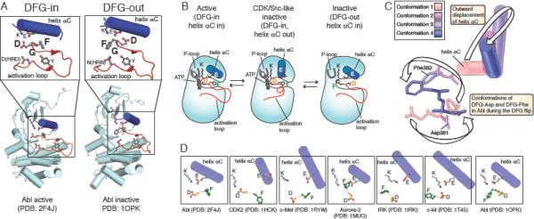
In contrast to the active conformations of protein kinases, which are essentially the same for all kinases, inactive kinase conformations are structurally diverse. Some inactive conformations are, however, observed repeatedly in different kinases, perhaps reflecting an important role in catalysis. In this review, we analyze one of these recurring conformations, first identified in CDK and Src kinases, which turned out to be central to understanding of how kinase domain of the EGF receptor is activated. This mechanism, which involves the stabilization of the active conformation of an α helix, has features in common with mechanisms operative in several other kinases.


Figures

Figure 1
Figure 1
Activation of protein kinases. (A) Crystal structure (PDB ID: 1ATP) and cartoon representation of the active state of the protein kinase A (PKA). The inset selectively displays the catalytically critical components of the kinase active site: the activation loop, helix αC, P-loop, ATP, magnesium ion and the catalytic residues: DFG-aspartate (Asp 184), HRD-aspartate (Asp 166), catalytic lysine (Lys 72) catalytic glutamate (Glu 91), and the autophosphorylation site in the activation loop (Thr 197). (B) The cartoon representation of the assembly of the hydrophobic spines (the regulatory and the catalytic spine) in the active state of a kinase and of their disassembly in the inactive state. The insets to the right of the cartoons represent surface representation of the residues corresponding to the regulatory and catalytic spines in the inactive Abl kinase (PDB ID: 1OPJ) and in the active Abl kinase (PDB ID: 2G2I). The DFG-phenylanine is depicted, as well as the catalytic lysine (K) and the catalytic glutamate in helix αC (E).
Figure 2
Figure 2
CDK/Src-like inactive conformation. (A) The activation mechanisms of the Src family of kinases and cyclin-dependent kinases (CDKs). When not bound to SH2 and SH3 domains, Src kinases adopt an active conformation. In contrast, CDKs are inactive when not bound to their regulators cyclins. Activation of CDKs requires cyclin binding. (B) Crystal structures of the Hck kinase (PDB ID: 1QCF) in the CDK/Src-like inactive conformation, Lck kinase (PDB ID: 3LCK) in the active conformation and the Hck kinase (PDB ID: 2HCK) in the alternate CDK/Src-like inactive conformation. For clarity the SH2 and SH3 domains are not shown in the inactive structures. The insets underneath the structures selectively display the activation loop, helix αC, P-loop and the selected residues to highlight major conformational changes between different states.
Figure 3
Figure 3
Coupling of the DFG flip to the CDK/Src-like inactive conformation. (A) Two distinct conformations of the DFG motif found in the crystal structures of the Abl kinase domain. In the active structure (PDB ID: 2F4J), DFG adopts the DFG-in conformation, in which the DFG-aspartate is positioned towards the active site. In the inactive, DFG-out conformation (PDB ID: 1OPK), DFG-phenylalanine flips towards the active site. The DFG residues are depicted as single letter amino acid codes (D, F, G), as is the catalytic lysine (K), the catalytic glutamate in helix αC (E) and the activation loop tyrosine (Y). (B) CDK/Src-like inactive conformation is proposed as an intermediate conformation adopted by kinases during DFG flipping. (C) A DFG-in to DFG-out flip observed in long time scale molecular dynamics simulations. Starting from conformation 1 (active Abl kinase crystal structure PDB: 2F4J), the DFG-aspartate migrates away from the active site, whereas DFG-phenylalanine enters from above. The accompanying displacement of helix αC is illustrated with conformations from different simulation times (figure adapted with permission from (Shan et al., 2009). (D) The series of kinase structures captures stepwise progression of the DFG flipping from the DFG-in to the DFG-out conformation, adapted with permission from (Shan et al., 2009). These steps are associated with the significant movement of helix αC and a transient adoption of the CDK/Src-like inactive conformation (PDB ID: IHCK (CDK2 kinase) and PDB ID: 1R1W (c-Met kinase)). The insets selectively highlight helix αC, the catalytic lysine (K), the catalytic glutamate (E) and the DFG-aspartate (D) and DFG-phenylalanine (F) residues.
Figure 4
Figure 4
The CDK/Src-like switch underlies the activation of the EGF receptor kinase domain. (A and B) Both CDKs and the EGF receptor (EGFR) kinase are stable in the inactive CDK/Src-like conformation due to a network of hydrophobic residues in the N-lobe (shown as dot representation in magenta color). During activation, the hydrophobic residues become exposed creating an interface (an activator-binding patch or a cyclin-binding patch) that binds allosteric activators of these kinases. In case of the EGF receptor (A), one kinase domain (the activator kinase) becomes an activator of the other (the receiver kinase) by forming a head to tail asymmetric dimer. CDKs are subject to the allosteric activation by cyclins (B).
Figure 5
Figure 5
Allosteric control of the EGF receptor dimerization. (A) Schematic representation of the EGF receptor domain organization. (B) The crystal structure and cartoon representation of the EGF receptor kinase (EGFR) domain in the presence of its full juxtamembrane segment (PDB ID: 3GOP) depicts binding of the juxtamembrane latch of the receiver kinase to the activator kinase. (C) The cartoon representation of the overlapping interactions involving the juxtamembrane latch-binding site, based on the structures of the active EGF receptor kinase domain dimer in the presence of its juxtamembrane segment (PDB ID: 3GOP), the inactive EGF receptor kinase dimer (PDB ID: 3GT8) and the inactive EGF receptor kinase bound to an inhibitor Mig6 (PDB ID: 2RFE). (D) The model for ligand-dependent activation of EGF receptor at the plasma membrane, which depicts contribution of the extracellular, transmembrane and the juxtamembrane domains to receptor dimerization.
Figure 6
Figure 6
HER3 receptor kinase domain is a specialized allosteric activator of other EGF receptor family kinases. (A) Cartoon representation of possible dimerization scenarios in the EGF receptor family of kinases. (B) The crystal structures of the inactive EGF receptor kinase domain (PDB ID: 3GT8) and the HER3 kinase domain (PDB ID: 3KEX). The HER3 kinase domain is in the CDK/Src-like inactive conformation and has altered conformation of helix αC.
Figure 7
Figure 7
A recurring mode for activation of kinases by engaging the helix αC patch. Crystal structures and cartoon representations of PKA (PDB ID: 1ATP), the Ret receptor kinase domain (PDB ID: 2IVT), the Aurora kinase (PDB ID: 1Ol5), the AKT/PKB kinase (PDB ID: 1O6K), the Rho kinase (PDB ID: 2V55) and the Fes kinase (PDB ID: 3CD3) depict different modes by which these kinases engage the helix αC patch (HM-binding pocket in AKT/PKB) during activation.

References

    1. Aifa S, Aydin J, Nordvall G, Lundstrom I, Svensson SP, Hermanson O. A basic peptide within the juxtamembrane region is required for EGF receptor dimerization. Exp Cell Res. 2005;302:108–114. - PubMed
    1. Azam M, Seeliger MA, Gray NS, Kuriyan J, Daley GQ. Activation of tyrosine kinases by mutation of the gatekeeper threonine. Nat Struct Mol Biol. 2008;15:1109–1118. - PMC - PubMed
    1. Bargmann CI, Hung MC, Weinberg RA. Multiple independent activations of the neu oncogene by a point mutation altering the transmembrane domain of p185. Cell. 1986a;45:649–657. - PubMed
    1. Bargmann CI, Hung MC, Weinberg RA. The neu oncogene encodes an epidermal growth factor receptor-related protein. Nature. 1986b;319:226–230. - PubMed
    1. Bayliss R, Sardon T, Vernos I, Conti E. Structural basis of Aurora-A activation by TPX2 at the mitotic spindle. Mol Cell. 2003;12:851–862. - PubMed

Publication types

MeSH terms