. 2011 Apr 8;42(1):50-61.
doi: 10.1016/j.molcel.2011.03.017.

Rac1 regulates the activity of mTORC1 and mTORC2 and controls cellular size

Affiliations

Rac1 regulates the activity of mTORC1 and mTORC2 and controls cellular size

Abdelhafid Saci et al. Mol Cell. .

Abstract

Mammalian target of rapamycin (mTOR) is a serine/threonine kinase that exists in two separate complexes, mTORC1 and mTORC2, that function to control cell size and growth in response to growth factors, nutrients, and cellular energy levels. Low molecular weight GTP-binding proteins of the Rheb and Rag families are key regulators of the mTORC1 complex, but regulation of mTORC2 is poorly understood. Here, we report that Rac1, a member of the Rho family of GTPases, is a critical regulator of both mTORC1 and mTORC2 in response to growth-factor stimulation. Deletion of Rac1 in primary cells using an inducible-Cre/Lox approach inhibits basal and growth-factor activation of both mTORC1 and mTORC2. Rac1 appears to bind directly to mTOR and to mediate mTORC1 and mTORC2 localization at specific membranes. Binding of Rac1 to mTOR does not depend on the GTP-bound state of Rac1, but on the integrity of its C-terminal domain. This function of Rac1 provides a means to regulate mTORC1 and mTORC2 simultaneously.


Figures

Figure 1
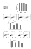
Figure 1. Rac1 Deletion Reduced the Size of MEFs and Lymphocytes
(A) Fl/+ and fl/fl MEFs were grown in 10% serum in the presence and absence of 4-HT for 48 hr (1: fl/+ treated with 4-HT, 2: fl/fl treated with ethanol, 3: fl/fl treated with 4-HT). Cells were lysed and subjected to SDS-PAGE and WB to assess Rac1 deletion. (B) Rac1 fl/+ and fl/fl MEFs were grown in 10% serum, resuspended in Trypsin, diluted in PBS, then subjected to cell size analysis on a Beckman Coulter cell sorter before and after 4-HT treatment. (C–F) B cells (C and D) and T cells (E and F) from WT, Rac1 fl/fl, Rac2−/− and Rac1 fl/fl Rac2−/− mice were grown in RPMI with 10 % serum for 48 hr in the presence of 500 nM 4-HT. Cells were resuspended in PBS, then subjected to FACscan analysis, based on forward scatter/side scatter (FS/SSC). The histograms in (D) and (F) resulted from the quantification of the cell size on the Forward Scatter scale. Error bars in (B), (D), and (F) specify the standard deviation between four independently performed experiments. Results are representative of four experiments.
Figure 2
Figure 2. Rac1 Regulates mTOR Activity in MEFs and HeLa Cells
(A and B) WT (A) and Rac1 fl/fl (B) MEFs were cultured in DMEM with 10% serum. They were treated with ethanol (control) or with 500 nM 4-HT for 48 hr to delete Rac1. In some conditions, cells were treated for 1 hr with 100 nM wortmannin or 100 nM rapamycin at 37°C. (C) Rac1 or control RNAi oligos were introduced into HeLa cells before analysis. (D) Immortalized MEFs were infected or not with control or Rac1-expressing lentiviral particles, then incubated or not with 4-HT for 48 hr with serum-starvation for the last 14 hr. They were then stimulated or not for 10 min with 10% serum supplemented with 50 nM PDGF. The cells were then lysed and subjected to SDS-PAGE and western blot analysis. These results are representative of three experiments.
Figure 3
Figure 3. Rac1 Regulates mTOR Activity Independently of PI3K, Binding to GTP/GDP and Actin Polymerization
(A) Primary Rac1 fl/fl MEFs were grown in 10% serum and treated or not with 4-HT for 48 hr, then serum starved (overnight). They were incubated or not with 100 nM wortmannin for 30 min then subjected to stimulation with 1 μM PMA for 15 min. (B) Cells were grown in 10% serum and treated with ethanol (control) or with 500 nM 4-HT for 48 hr to delete Rac1. In some conditions, cells were treated for 1 hr with 100 nM wortmannin or 100 nM Rapamycin at 37°C or with 10 μM of a Rac inhibitor, NSC-23766. (C) MEFs were treated or not with 500 nM 4-HT for 36 hr in DMEM with 10% heat-inactivated FBS to delete Rac1. Cells were then infected with recombinant vaccinia viruses, expressing Rac1 WT, V12Rac1, N17Rac1, or the empty vector pSC65 for 14 hr. (D) HeLa cells were grown in 10% serum then serum starved for 14 hr. They were treated or not with 10 μg/ml cytochalasin D and stimulated or not with 10% serum supplemented with 50 nM PDGF. Cells were then lysed and subjected to SDS-PAGE and western blot analysis. These results are representative of three experiments.
Figure 4
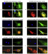
Figure 4. Rac1 Is Colocalized with mTOR and Is Required for Subcellular Localization of mTOR
(A and B) MEFs were serum-starved (a) or serum starved then stimulated with 10% serum for 10 min (B). Cells were fixed and immunostained for Rac1 and mTOR and analyzed by immunofluorescence. (C and D) MEFs were serum starved (C) or serum starved then stimulated with 10% serum, supplemented with 50 nM PDGF for 10 min (D). They were fixed and immunostained for Rac1 and mTOR and subjected to confocal microscopy analysis as described in the method section. (E and F) Rac1 fl/fl MEFs were incubated with 4-HT for 2 days. They were serum starved (E) and serum-starved then stimulated for 10 min (F). Cells were fixed and stained for Rac1 and mTOR. Cells were visualized by immunofluorescence as described in the methods section. Images are representative of four experiments.
Figure 5
Figure 5. Rac1 Is Colocalized with Both mTORC1 and mTORC2 Components
(A–C) HeLa cells were serum starved then stimulated with 10% serum for 20 min. They were fixed and immunostained for Rac1 and Raptor (A) or Rac1 and Rictor (B) or Rac1 and Sin1 (C), then analyzed by immunofluorescence as described in the Experimental Procedures section. Images are representative of four experiments.
Figure 6
Figure 6. Rac1 Interacts with mTORC1 and mTORC2 Proteins
(A) Fifty to sixty percent confluent 293T cells were treated with PolyEthylImine (PEI) as a control or transfected with HA-tagged Rac1 or Rac2 and the different Myc-tagged mTORC proteins using PEI, as indicated. (B) 293T cells were treated with PEI (control) or transfected with HA-tagged WT Rac1, V12Rac1, or N17Rac1 mutants and with either mTOR, Ric tor, or Raptor as indicated. Cells were then lysed and subjected to HA IP. (C) 293T cells were transfected with either mTOR, Rictor or Raptor. Cells were lysed and subjected to pull-down assays using GST-fusion proteins of WT and Rac1 mutants for 1 hr at 4°C. The protein complexes were washed and then analyzed by SDS-PAGE and western blot. Results are representative of three experiments. (D) 293T cells were transfected with either mTOR, Rictor, or Raptor. Cells were lysed and subjected to pull-down assays using GST-fusion proteins of WT and Rac1 mutants for 1 hr at 4°C. The protein complexes were washed and then analyzed by SDS–PAGE and western blot. Results are representative of three experiments.
Figure 7
Figure 7. The C-Terminal Region of Rac1 Is Required for the Direct Interaction with mTOR
(A) Fifty to sixty percent confluent 293T cells were treated with PEI (control) or transfected with mTOR, Rictor, or Raptor. Cells were then lysed and biotin-peptides of the Rac1, Rac2, and Cdc42 C-terminal domains and mutated Rac1 peptide: PPP/AAA or RKR/AAA was used to precipitate mTORC proteins for 1 hr at 4°C in the presence of streptavidin beads. (B and C) 293T cells were transfected with Flag-mTOR. The Flag IPs were eluted by the Flag peptide. The free mTOR protein was precipitated by biotin-Rac1 C-terminal peptide or the biotin-RKR/AAA mutant, followed by streptavidin agarose. Samples were analyzed by SDS-PAGE and western blot (b) or the SDS-PAGE was silver stained (C). MW: molecular weight markers, WL: total proteins from Flag-mTOR transfected cells (1) WT biotin-Rac1 peptide; (2) biotin RKR/AAA mutant; (3) Flag IP. Data are representative of three experiments.

Comment in

  • Cell growth: RAC1 sizes up mTOR.
    Schuldt A. Schuldt A. Nat Rev Mol Cell Biol. 2011 Jun;12(6):343. doi: 10.1038/nrm3124. Epub 2011 May 18. Nat Rev Mol Cell Biol. 2011. PMID: 21587295 No abstract available.

References

    1. Avruch J, Hara K, Lin Y, Liu M, Long X, Ortiz-Vega S, Yonezawa K. Insulin and amino-acid regulation of mTOR signaling and kinase activity through the Rheb GTPase. Oncogene. 2006;25:6361–6372. - PubMed
    1. Bain J, Plater L, Elliott M, Shpiro N, Hastie CJ, McLauchlan H, Klevernic I, Arthur JS, Alessi DR, Cohen P. The selectivity of protein kinase inhibitors: a further update. Biochem. J. 2007;408:297–315. - PMC - PubMed
    1. Benvenuti F, Hugues S, Walmsley M, Ruf S, Fetler L, Popoff M, Tybulewicz VL, Amigorena S. Requirement of Rac1 and Rac2 expression by mature dendritic cells for T cell priming. Science. 2004;305:1150–1153. - PubMed
    1. Berven LA, Willard FS, Crouch MF. Role of the p70(S6K) pathway in regulating the actin cytoskeleton and cell migration. Exp. Cell Res. 2004;296:183–195. - PubMed
    1. Burridge K, Wennerberg K. Rho and Rac take center stage. Cell. 2004;116:167–179. - PubMed

Publication types

MeSH terms