. 2011 Apr 8;42(1):84-95.
doi: 10.1016/j.molcel.2011.01.030.

Integrating cardiac PIP3 and cAMP signaling through a PKA anchoring function of p110γ

Affiliations

Integrating cardiac PIP3 and cAMP signaling through a PKA anchoring function of p110γ

Alessia Perino et al. Mol Cell. .

Abstract

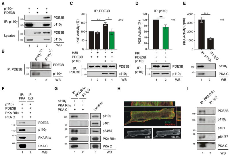
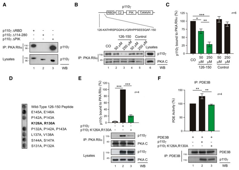
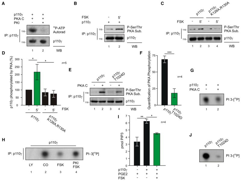
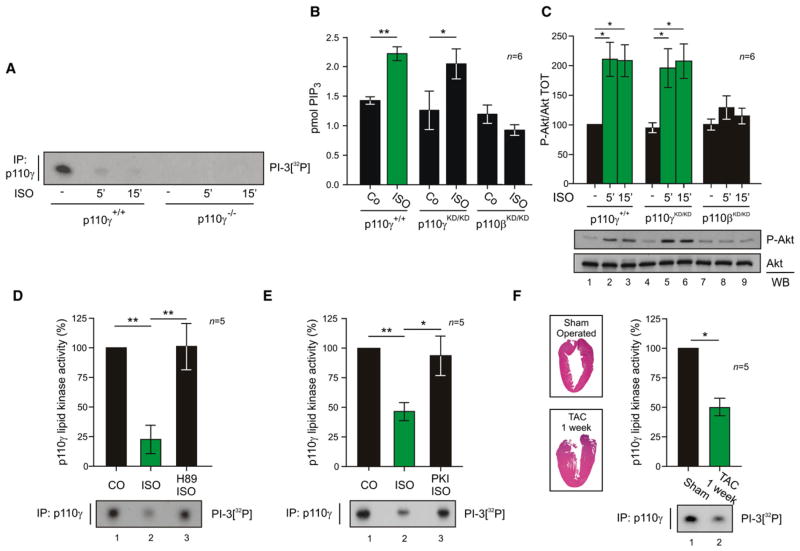
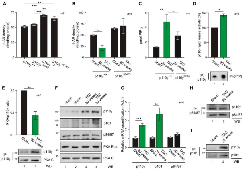
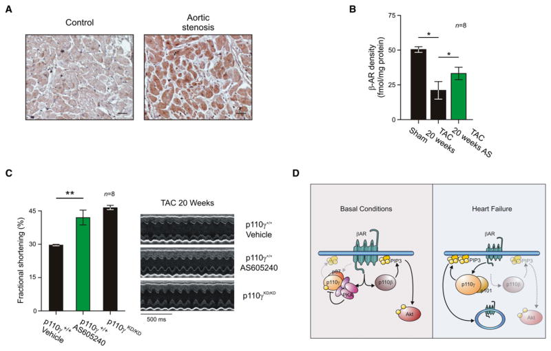
Adrenergic stimulation of the heart engages cAMP and phosphoinositide second messenger signaling cascades. Cardiac phosphoinositide 3-kinase p110γ participates in these processes by sustaining β-adrenergic receptor internalization through its catalytic function and by controlling phosphodiesterase 3B (PDE3B) activity via an unknown kinase-independent mechanism. We have discovered that p110γ anchors protein kinase A (PKA) through a site in its N-terminal region. Anchored PKA activates PDE3B to enhance cAMP degradation and phosphorylates p110γ to inhibit PIP(3) production. This provides local feedback control of PIP(3) and cAMP signaling events. In congestive heart failure, p110γ is upregulated and escapes PKA-mediated inhibition, contributing to a reduction in β-adrenergic receptor density. Pharmacological inhibition of p110γ normalizes β-adrenergic receptor density and improves contractility in failing hearts.


Figures

Figure 1
Figure 1. p110γ Activates PDE3B through PKA within a PI3Kγ-PKA-PDE3B Complex
(A) Coimmunoprecipitation of PDE3B with p110γ in HEK293T cells transfected with p110γ and PDE3B-Flag. (B) Coimmunoprecipitation of p110γ with PDE3B in wild-type (p110γ+/+) but not in p110γ knockout (p110γ−/−) mouse neonatal cardiomyocytes. (C) Phosphodiesterase activity in PDE3B immunoprecipitates upon transfection of HEK293T cells with PDE3B-Flag (PDE3B) or with PDE3B-Flag and p110γ. Cells were treated with PKA inhibitor H89 (5 μM, 10 min) or vehicle as indicated. PDE activity (%) was calculated relative to the activity of single PDE3B transfectants. (D) Phosphodiesterase activity (%) of double p110γ, PDE3B-Flag transfectants treated with PKA inhibitor Myr-PKI (5 u;M, 10 min) or vehicle. (E) PKA activity (cpm) in a p110γ immunoprecipitate from transfected HEK293T cells. (F) Coimmunoprecipitation of transfected p110γ, PDE3B-Flag, PKA RIIα-ECFP (PKA RIIα), and PKA CAT-YFP (PKA C) from HEK293T extracts. (G) Coimmunoprecipitation of p110γ and p84/p87, but not p101, with PKA RIIα from HEK293T transfected cells. (H) Colocalization (yellow spots) of p110γ (green) and PKA RIIα (red) by immunofluorescence in mouse adult cardiomyocytes. Longitudinal and transverse sections are shown in the upper and right panels, respectively. Single p110γ and PKA RIIα localizations are presented in the lower panels. (I) PDE3B, p110γ, p84/p87, and PKA CAT (PKA C), but not p101, coimmunoprecipitate with PKA RIIα in myocardial tissue extracts of wild-type mice. A representative immunoprecipitation is presented in (A)–(G) and (I). For all bar graphs, values represent mean ± SEM of a minimum of four independent experiments. *p < 0.05, **p < 0.01, ***p < 0.001. See also Figures S1 and S2.
Figure 2
Figure 2. p110γ Is a Bona Fide AKAP
(A) In vitro copurification of p110γ-GST and PKA RIIα-6His in a GST pull-down assay. (B) Direct interaction of p110γ and PKA RIIα in a surface plasmon resonance assay. p110γ was immobilized on the chip, and PKA RIIα was injected at three different concentrations. (C) Binding of 32P-labeled PKA RIIα to immunoprecipitated p110γ in the presence of AKAP-IS scrambled peptide but not in the presence of AKAP-IS peptide. (D) Competition of the p110γ-PKA RIIα coimmunoprecipitation with AKAP-IS peptide. (E) Quantitative densitometry of the competition experiment represented in (D). Values represent mean ± SEM of four independent experiments. *p < 0.05. (F) Loss of the coimmunoprecipitation of PKA RIIα with p110γ by truncation of the 1–45 amino acids of RIIα-ECFP (PKA RIIα Δ1–45). A representative assay is presented in all figures. See also Figure S3.
Figure 3
Figure 3. Mapping of the p110γ-PKA RIIα Interaction
(A) Loss of the coimmunoprecipitation of p110γ-Myc with PKA RIIα by deletion of amino acids 114–280 (p110γ Δ 114–280) but not by deletion of the Ras-binding domain (p110γ Δ RBD) or the PIK domain (p110γ Δ PIK) of p110γ-Myc. (B) Sequence of the 126–150 peptide is represented on a scheme of p110γ. In dose-dependent competition experiments, the 126–150 peptide, but not a scrambled control peptide, antagonized the p110γ-RIIα protein-protein interaction. (C) Quantitative densitometry of the competition experiment represented in (B). (D) Binding of PKA RIIα to a set of alanine mutant 126–150 p110γ peptides in a solid-phase peptide array. (E) Mutation of K126 and R130 of p110γ to A (p110γ K126A, R130A) blunts coimmunoprecipitation of p110γ with PKA RIIα in transfected HEK293T cells. Values were obtained by quantitative densitometry and normalized over control. (F) Phosphodiesterase activity (%) of PDE3B immunoprecipitates upon transfection of HEK293T cells with PDE3B-Flag alone or with PDE3B-Flag and either wild-type or mutant p110γ (p110γ K126A, R130A). A representative immunoprecipitation is provided for (A), (B), (E), and (F). For all bar graphs, values represent mean ± SEM of a minimum of four independent experiments. *p < 0.05, **p < 0.01, ***p < 0.001. See also Figure S4.
Figure 4
Figure 4. PKA Phosphorylates and Inhibits p110γ
(A) Phosphorylation of p110γ immunoprecipitated from transfected HEK293T cells in the presence of recombinant PKA C, 32P-ATP, and PKA inhibitor PKI (1 μM) or vehicle. (B) PKA-mediated phosphorylation of p110γ immunoprecipitated from HEK293T cells upon stimulation with forskolin (FSK 20 μM, 5 min). (C) PKA-mediated phosphorylation of p110γ or p110γ K126A, R130A immunoprecipitated from HEK293T cells upon stimulation with forskolin (FSK 20 μM, 5 min). (D) Quantitative densitometry of the experiment represented in (C). (E) PKA-mediated phosphorylation of p110γ or p110γ T1024D mutant immunoprecipitated from HEK293T cells and incubated or not with active PKA (30 min). (F) Quantitative densitometry of PKA phosphorylation (background subtracted) of the experiment represented in (E). (G) Lipid kinase activity of recombinant p110γ-GST incubated in vitro with or without recombinant PKA C (30 min). (H) Lipid kinase activity of p110γ immunoprecipitated from transfected HEK293T cells treated with vehicle, pan-p110 inhibitor LY-294002 (LY, 20 μM, 15 min), forskolin (FSK, 20 μM, 5 min) or FSK plus PKA inhibitor Myr-PKI (1 μM). (I) Measurement of cellular PtdIns(3,4,5)P3 (pmol PIP3) levels in transfected HEK293T treated with the GPCR agonist PGE2 (100 nM, 10 min) alone or in combination with FSK (50 μM, 3 min). (J) Lipid kinase activity of p110γ wild-type or p110γ T1024D mutant immunoprecipitated from transfected HEK293T cells. In lipid kinase assays (G, H, J), the ability of p110γ to phosphorylate phosphoinositide was detected by autoradiography following incubation with 32P-ATP substrate. A representative assay is presented in all figures. For all bar graphs, values represent mean ± SEM of a minimum of four independent experiments. *p < 0.05, **p < 0.01, ***p < 0.001. See also Figures S5 and S6.
Figure 5
Figure 5. PKA Inhibits the Lipid Kinase Activity of p110γ in the Myocardium
(A) Lipid kinase activity of p110γ immunoprecipitated from myocardial tissue lysates following treatment of wild-type (p110γ+/+) or p110γ knockout mice (p110γ−/−) with β-AR agonist isoproterenol (ISO, 1.25 mg/kg i.p. for 5 or 15 min) or vehicle. (B) Measurement of myocardial PtdIns(3,4,5)P3 (pmol PIP3) levels in wild-type (p110γ+/+), p110γ kinase-dead (p110γKD/KD), and p110β kinase-dead (p110βKD/KD) mice treated with isoproterenol (ISO, 1.25 mg/kg i.p. for 5 min) or vehicle. (C) Phospho-Akt (P-Akt) and total Akt (Akt) levels in myocardial tissue lysates following treatment of wild-type (p110γ+/+), p110γ kinase-dead (p110γKD/KD), and p110β kinase-dead (p110βKD/KD) mice with β-AR agonist isoproterenol (ISO, 1.25 mg/kg i.p. for 5 or 15 min) or vehicle. (D) Lipid kinase activity of p110γ immunoprecipitated from myocardial tissue lysates following ex vivo cardiac Langendorff perfusion (5 min) with vehicle, isoproterenol (ISO 10 μM), or ISO plus PKA inhibitor H89 (10 μM). (E) Lipid kinase activity of p110γ immunoprecipitated from rat adult cardiomyocytes treated with isoproterenol (ISO, 1 μM, 3 min), ISO plus PKA inhibitor Myr-PKI (1 μM, 5 min), or vehicle. (F) Lipid kinase activity of p110γ immunoprecipitated from myocardial tissue lysates of mice subjected to transverse aortic constriction for 1 week (TAC 1 week) or to sham operation. Insets are representative hematoxylin and eosin stainings of left ventricular sections from sham-operated and 1 week TAC mice. A representative assay is presented in all figures. For all bar graphs, values, obtained by quantitative densitometry and normalized over control, represent mean ± SEM of a minimum of five mice per group. *p < 0.05, **p < 0.01.
Figure 6
Figure 6. Modulation of p110γ Lipid Kinase Activity by PKA Affects β-AR Density
(A) Myocardial β-AR density in wild-type (p110γ+/+), p110β kinase-dead (p110βKD/KD), p110γ knockout (p110γ−/−), and p110γ kinase-dead (p110γKD/KD) mice. (B) Myocardial β-AR density of p110γ+/+ and p110γKD/KD mice subjected to sham operation or to aortic constriction for 20 weeks (TAC 20 weeks). (C) Myocardial PtdIns(3,4,5)P3 (pmol PIP3) levels in hearts obtained from sham-operated mice or from 20 week TAC-treated wild-type or p110γKD/KD mice. (D) Lipid kinase activity of p110γ immunoprecipitated from myocardial tissue lysates of sham-operated or 20 week TAC-treated mice. Values were obtained by quantitative densitometry and normalized over control. (E) Coimmunoprecipitation of p110γ and PKA C from myocardial lysates from mice subjected to pressure overload for 20 weeks or to sham operation. After quantitative densitometry, p110γ-bound PKA was expressed as the ratio of coimmunoprecipitated PKA C over immunoprecipitated p110γ. (F) Total levels of the indicated proteins in myocardial tissue lysates from sham-operated mice or from mice subjected to pressure overload for 20 weeks. (G) mRNA levels of p110γ, p101, and p84/p87 in hearts from sham-operated mice or mice subjected to 20 weeks of TAC. (H) Coimmunoprecipitation of p110γ with p84/87 from myocardial lysates of mice subjected to pressure overload for 20 weeks or to sham operation. (I) Coimmunoprecipitation of p110γ with p101 from myocardial lysates of mice subjected to pressure overload for 20 weeks or to sham operation. In β-AR density measurements (A and B), receptor density is expressed as the Bmax after saturation binding using 125I-labeled cyanopindolol ligand (fmol/mg protein). A representative assay is presented in (D)–(I). For all bar graphs, values represent mean ± SEM of a minimum of four independent experiments or six mice per group. *p < 0.05, **p < 0.01, ***p < 0.001. See also Figure S7.
Figure 7
Figure 7. p110γ Inhibition Restores β-AR Density and Cardiac Function in Heart Failure
(A) Immunohistochemistry for p110γ in human hearts from healthy control patients or patients with aortic stenosis. Bar graph is 25 μm. Representative images are presented. (B) Myocardial β-AR density of wild-type mice with heart failure (obtained by 20 weeks of pressure overload) treated with selective p110γ inhibitor AS605240 (AS 10 mg/kg i.p. q.d. for 1 week) or vehicle. (C) Left ventricular fractional shortening of wild-type mice with heart failure treated with AS605240 (10 mg/kg i.p. q.d. for 1 week) or vehicle or of p110γKD/KD mice subjected to aortic constriction for 20 weeks. Representative M-mode echocardiographic snapshots are presented (right). For all bar graphs, values represent mean ± SEM of eight mice per group. *p < 0.05, **p < 0.01. (D) Schematic representation of the diverse function of p110γ and p110β in cardiac physiology and pathology. See also Figure S7.

References

    1. Alto NM, Soderling SH, Hoshi N, Langeberg LK, Fayos R, Jennings PA, Scott JD. Bioinformatic design of A-kinase anchoring protein-in silico: a potent and selective peptide antagonist of type II protein kinase A anchoring. Proc Natl Acad Sci USA. 2003;100:4445–4450. - PMC - PubMed
    1. Bristow MR, Ginsburg R, Minobe W, Cubicciotti RS, Sageman WS, Lurie K, Billingham ME, Harrison DC, Stinson EB. Decreased catecholamine sensitivity and beta-adrenergic-receptor density in failing human hearts. N Engl J Med. 1982;307:205–211. - PubMed
    1. Bristow MR, Hershberger RE, Port JD, Gilbert EM, Sandoval A, Rasmussen R, Cates AE, Feldman AM. Beta-adrenergic pathways in nonfailing and failing human ventricular myocardium. Circulation. 1990;82 (2 Suppl):I12–I25. - PubMed
    1. Carr DW, Stofko-Hahn RE, Fraser ID, Bishop SM, Acott TS, Brennan RG, Scott JD. Interaction of the regulatory subunit (RII) of cAMP-dependent protein kinase with RII-anchoring proteins occurs through an amphipathic helix binding motif. J Biol Chem. 1991;266:14188–14192. - PubMed
    1. Ciccarelli M, Santulli G, Campanile A, Galasso G, Cervèro P, Altobelli GG, Cimini V, Pastore L, Piscione F, Trimarco B, Iaccarino G. Endothelial alpha1-adrenoceptors regulate neo-angiogenesis. Br J Pharmacol. 2008;153:936–946. - PMC - PubMed

Publication types

MeSH terms

Substances