. 2011 Apr 8;8(4):376-88.
doi: 10.1016/j.stem.2011.03.001.

Highly efficient miRNA-mediated reprogramming of mouse and human somatic cells to pluripotency

Affiliations

Highly efficient miRNA-mediated reprogramming of mouse and human somatic cells to pluripotency

Frederick Anokye-Danso et al. Cell Stem Cell. .

Erratum in

  • Cell Stem Cell. 2012 Dec 7;11(6):853

Abstract

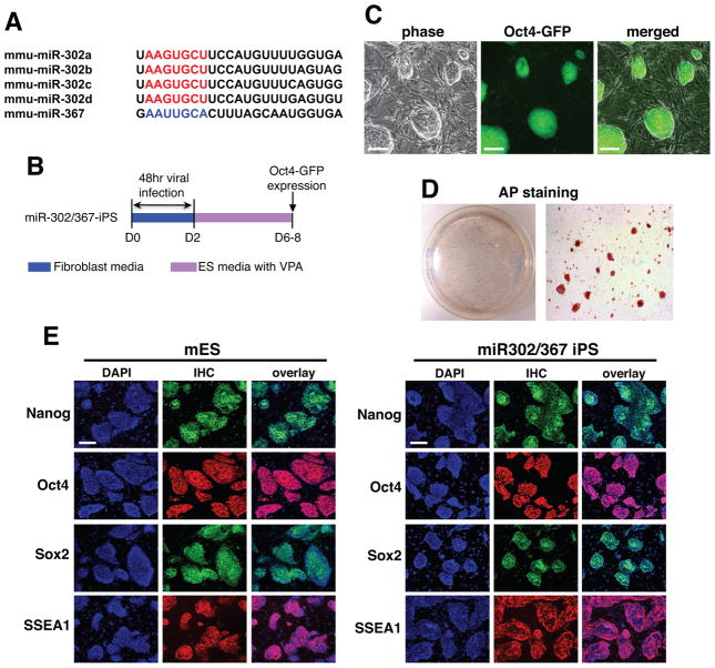
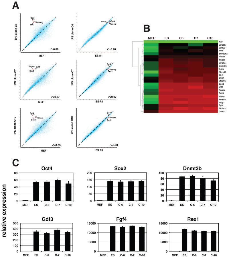
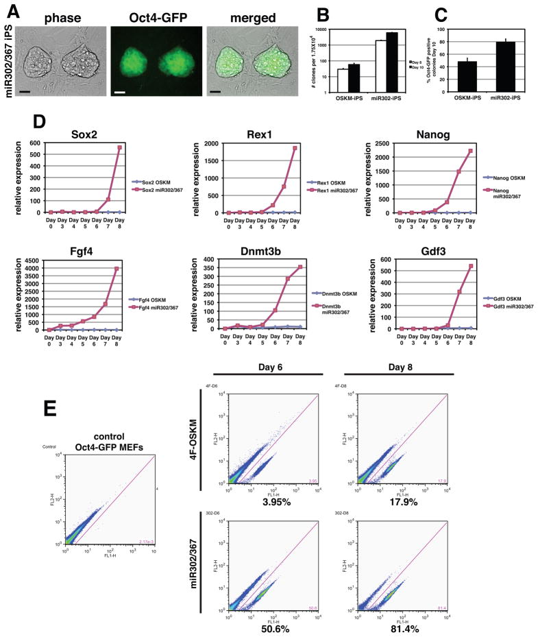
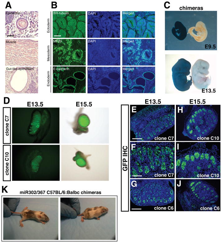
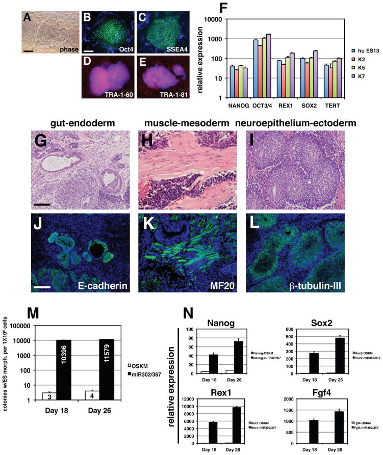
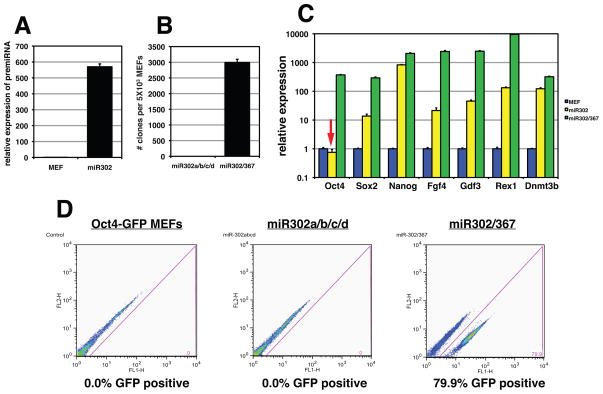
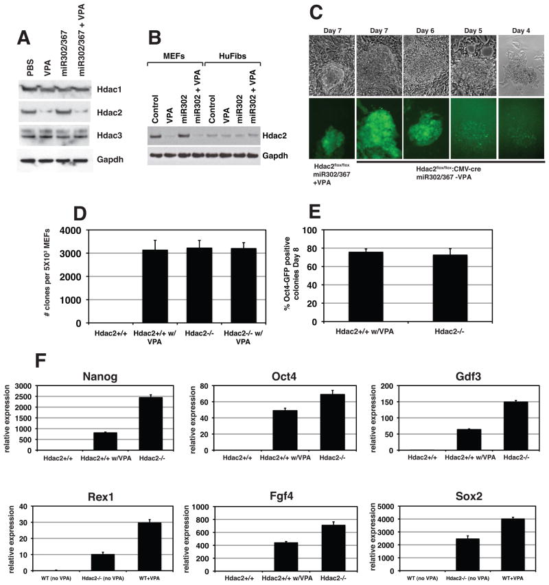
Transcription factor-based cellular reprogramming has opened the way to converting somatic cells to a pluripotent state, but has faced limitations resulting from the requirement for transcription factors and the relative inefficiency of the process. We show here that expression of the miR302/367 cluster rapidly and efficiently reprograms mouse and human somatic cells to an iPSC state without a requirement for exogenous transcription factors. This miRNA-based reprogramming approach is two orders of magnitude more efficient than standard Oct4/Sox2/Klf4/Myc-mediated methods. Mouse and human miR302/367 iPSCs display similar characteristics to Oct4/Sox2/Klf4/Myc-iPSCs, including pluripotency marker expression, teratoma formation, and, for mouse cells, chimera contribution and germline contribution. We found that miR367 expression is required for miR302/367-mediated reprogramming and activates Oct4 gene expression, and that suppression of Hdac2 is also required. Thus, our data show that miRNA and Hdac-mediated pathways can cooperate in a powerful way to reprogram somatic cells to pluripotency.


Figures

Figure 1
Figure 1. miR302/367 can reprogram mouse fibroblasts to a pluripotent stem cell phenotype
(A) The sequences of the miR302/367 cluster showing the similarity between members of the miR302a/b/c/d subfamily. miR367 has a different seed sequence than miR302a/b/c/d. (B) Schematic of viral expression protocol for miR302/367 iPS reprogramming with VPA. Day 0 is the start of viral transduction. (C) Oct4-GFP positive miR302/367 clones at seven days after starting viral transduction. (D) AP staining of a primary induction plate of miR302/367 iPS clones at eight days after starting viral transduction. (E) Immunostaining for Nanog, Oct4, Sox2, and SSEA1 in both mouse ES and primary induction samples of miR302/367 iPS cells at day 10 showing expression of pluripotent genes. See also Figures S1 and S2. Scale bars=100 μm.
Figure 2
Figure 2. miR302/367 iPSC clones have a similar expression profile as mouse ES cells
(A) Microarray experiments were used to show the similarity between miR302/367 iPS cell clones C6, C7, and C10 at passage 15 and the mouse ES cell line R1. (B) Heatmap of pluripotent gene expression of mouse ES cell line R1 and miR302/367 iPS cell clones C6, C7, and C10 from experiment in A. (C) Q-PCR of pluripotent gene expression of miR302/367 iPS cell clones C6, C7, and C10 at and mouse ES cell line R1. See also Figures S1 and S2.
Figure 3
Figure 3. miR302/367 plus VPA is two orders of magnitude more efficient that OSKM factors in iPS reprogramming of mouse fibroblasts
(A) miR302/367 iPS clones are readily observed 6–7 days after starting viral transduction and express high levels of Oct4-GFP while OSKM induced clones are not observed until 8–10 days, are very rare, and do not express significant levels of GFP from the Oct4 locus. (B) Counts of clones with ES like morphology from transduction of 1.75 × 10⁴ Oct4-GFP MEFs with equivalent amounts of either OSKM or miR302/367 virus at eight and ten days after viral transduction. Data are the average of three assays ± S.E.M. (C) Percentage of Oct4-GFP positive clones ten days after viral transduction with OSKM or miR302/367. Data are the average of three assays ± S.E.M. (D) Q-PCR of the indicated pluripotent factors comparing OSKM versus miR302/367 during the first eight days after viral transduction. (E) FACS analysis of miR302/367 reprogrammed Oct4-GFP MEFs compared to OSKM reprogrammed MEFs at six and eight days post-viral transduction. Scale bars=50 μm.
Figure 4
Figure 4. miR302/367 iPS cells can generate derivatives of mesoderm, endoderm, and ectoderm and contributue to the germline of mice
(A) Hematoxylin and eosin staining of teratomas derived from miR302/367 iPS cell clones showing skin epidermal-like structures, muscle, and gut-like epithelium. These data are representative of five different miR302/367 iPS cell clones which were injected and all produced teratomas. (B) Immunostaining of miR302/367 iPS derived teratoma tissues showing expressing of βIII-tubulin positive neural epithelium, MF20 positive striated muscle, and E-cadherin positive endodermal cells. (C) miR302/367 iPS clones can generate all tissues within the developing embryo as shown by lacZ histochemical staining of high percentage chimeric embryos derived from Rosa26-miR302/367 iPS clones at both E9.5 and E13.5. (D) Both whole mount fluorescence (D) and immunostaining for Oct4-GFP protein expression (E–J) show high-level contribution of miR302/367 iPS cell clones to the germline within the gonads of recipient mice. The data are representative of three clones (C6, C7, C10) which were injected into blastocysts and all three contributed to the germline. (K) miR302/367 iPSCs generated from C57BL/6 MEFs generate high percentage postnatal chimeras as noted by coat color. See also Figure S3. Scale bars: A=100 μm and B, D, G, H, J=150 μm, F and I=100 μm.
Figure 5
Figure 5. miR302/367 reprograms human fibroblasts to a pluripotent state more efficiently than OSKM factors
(A–E) Colony morphology and OCT4, SSEA4, TRA-1-60, and TRA-1-81 immunostaining of miR302/367 reprogrammed human fibroblasts. (F) Q-PCR of pluripotent stem cell marker genes in three different miR302/367 reprogrammed human fibroblast lines as compared to the human ES line HUES13. (G–I) Hematoxylin and eosin staining of teratomas derived from miR302/367 human iPS cell clones showing endoderm (gut), mesoderm (muscle), and ectoderm (neural epithelium) like structures. These data represent the results from seven human miR302/367 iPS cell clones. (J–L) Immunostaining of miR302/367 human iPS cell derived teratoma tissues showing expressing of E-cadherin positive endodermal cells, MF20 positive striated muscle, and βIII-tubulin positive neural epithelium. (M) Efficiency of miR302/367 reprogramming in human foreskin fibroblasts by colony counts of clones with human ES like morphology at 18 and 26 days post-viral transduction. Data are the average of three assays ± S.E.M. (N) Q-PCR of pluripotent gene expression in miR302/367 reprogrammed human foreskin fibroblasts at 18 and 26 days post-viral transduction. Data are the average of three assays ± S.E.M. See also Figures S1, S2, and S4. Scale bars: A–E= 50 μm, G–L=150 μm.
Figure 6
Figure 6. miR367 expression is required for miR302/367 iPS cell reprogramming
(A) The miR302a/b/c/d pre-miRNA is expressed at high levels in transduced MEFs. (B) Number of colonies generated after 10 days of miR302a/b/c/d or miR302/367 expression. Data are the average of four assays ± S.E.M. (C) Pluripotent gene expression from primary induction plates eights days after viral induction of miR302a/b/c/d or miR302/367 viruses. Note lack of Oct4 gene expression in miR302a/b/c/d expressing cells (red arrow). Data are the average of three assays ± S.E.M. (D) FACS analysis of Oct4-GFP MEFs eight days after transduction with either miR302a/b/c/d or miR302/367 viruses.
Figure 7
Figure 7. VPA specifically degrades Hdac2 protein and suppression of Hdac2 is required for iPS reprogramming by miR302/367
(A) VPA specifically degrades Hdac2 but not Hdac1 or Hdac3 proteins. Expression of miR302/367 alone did not have any affect on Hdac1, −2, or −3 protein levels. (B) Human foreskin fibroblasts express much lower levels of Hdac2 than MEFs. (C) Hdac2−/− MEFs start to reprogram between six and seven days post viral transduction which is similar to wild-type MEFs treated with VPA. (D) Number of clones generated with Hdac2−/− MEFs in the absence of VPA is similar to Hdac2+/+ MEFs with VPA at eight days post-viral transduction. Hdac2+/+ MEFs without VPA treatment did not generate any viable clones and VPA addition to Hdac2−/− MEFs did not increase the number of clones generated. (E) Percentage of Oct4-GFP positive clones is similar for Hdac2+/+ MEFs with VPA treatment and Hdac2−/− MEFs without VPA treatment at eight days post-viral transduction. (F) Q-PCR for pluripotent stem cell marker genes shows enhanced expression of pluripotency markers at day eight of reprogramming by miR302/367 in wild-type and Hdac2−/− MEFs versus Hdac2+/+ MEFs without VPA treatment. Data are the average of three assays ± S.E.M. See also Figures S5 and S6.

Comment in

Similar articles

Cited by

References

    1. Babiarz JE, Ruby JG, Wang Y, Bartel DP, Blelloch R. Mouse ES cells express endogenous shRNAs, siRNAs, and other Microprocessor-independent, Dicer-dependent small RNAs. Genes Dev. 2008;22:2773–2785. - PMC - PubMed
    1. Betel D, Wilson M, Gabow A, Marks DS, Sander C. The microRNA.org resource: targets and expression. Nucleic Acids Res. 2008;36:D149–153. - PMC - PubMed
    1. Bhutani N, Brady JJ, Damian M, Sacco A, Corbel SY, Blau HM. Reprogramming towards pluripotency requires AID-dependent DNA demethylation. Nature. 2010;463:1042–1047. - PMC - PubMed
    1. Blelloch R, Venere M, Yen J, Ramalho-Santos M. Generation of induced pluripotent stem cells in the absence of drug selection. Cell Stem Cell. 2007;1:245–247. - PMC - PubMed
    1. Card DA, Hebbar PB, Li L, Trotter KW, Komatsu Y, Mishina Y, Archer TK. Oct4/Sox2-regulated miR-302 targets cyclin D1 in human embryonic stem cells. Mol Cell Biol. 2008;28:6426–6438. - PMC - PubMed

Publication types