. 2011 Apr 8;8(4):389-98.
doi: 10.1016/j.stem.2011.02.002.

Bone marrow-derived cell therapy stimulates endogenous cardiomyocyte progenitors and promotes cardiac repair

Affiliations

Bone marrow-derived cell therapy stimulates endogenous cardiomyocyte progenitors and promotes cardiac repair

Francesco S Loffredo et al. Cell Stem Cell. .

Erratum in

  • Cell Stem Cell. 2015 Jul 2;17(1):125

Abstract

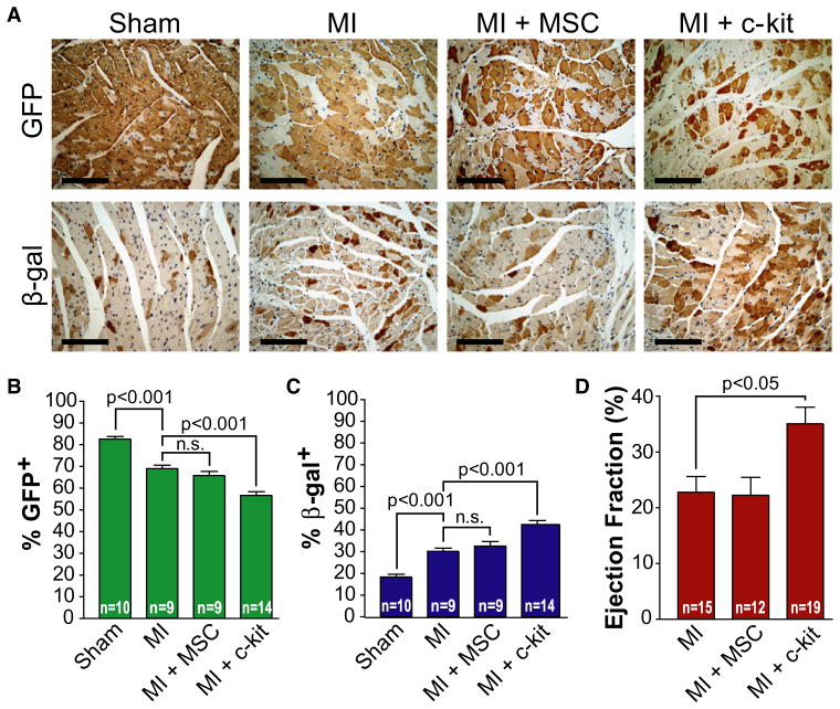
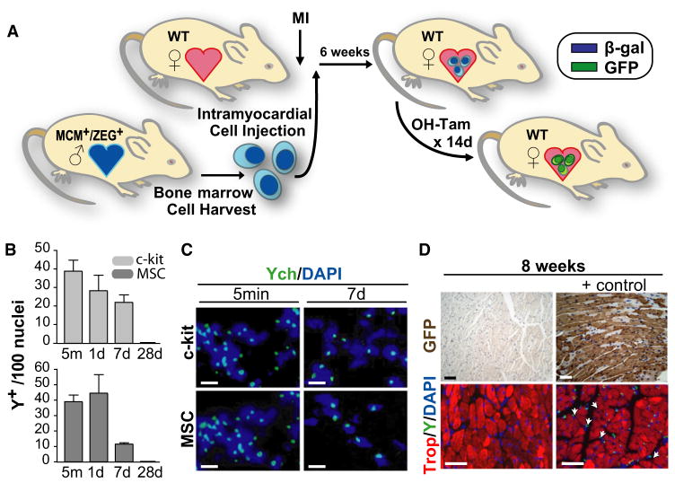
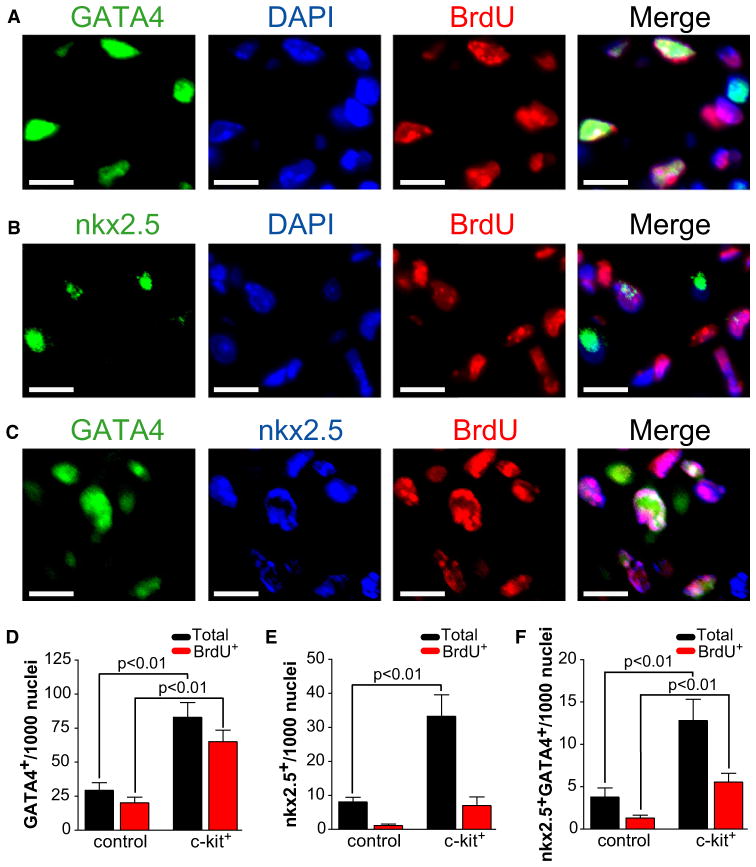
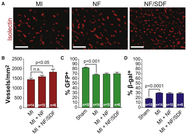
Cell therapy can improve cardiac function in animals and humans after injury, but the mechanism is unclear. We performed cell therapy experiments in genetically engineered mice that permanently express green fluorescent protein (GFP) only in cardiomyocytes after a pulse of 4-OH-tamoxifen. Myocardial infarction diluted the GFP(+) cardiomyocyte pool, indicating refreshment by non-GFP(+) progenitors. Cell therapy with bone marrow-derived c-kit(+) cells, but not mesenchymal stem cells, further diluted the GFP(+) pool, consistent with c-kit(+) cell-mediated augmentation of cardiomyocyte progenitor activity. This effect could not be explained by transdifferentiation to cardiomyocytes by exogenously delivered c-kit(+) cells or by cell fusion. Therapy with c-kit(+) cells but not mesenchymal stem cells improved cardiac function. These findings suggest that stimulation of endogenous cardiogenic progenitor activity is a critical mechanism of cardiac cell therapy.


Figures

Figure 1
Figure 1. Intramyocardial Delivery of Bone Marrow-Derived c-kit+ Cells after Myocardial Infarction Stimulates Endogenous Cardiomyocyte Regeneration
(A) Experimental protocol. MCM+/ZEG+ bitransgenic female mice were treated for 2 weeks with 4-OH-tamoxifen (OH-Tam) to induce a cardiomyocyte-specific switch from β-galactosidase (β-gal) to GFP expression. Experimental MI was performed by coronary ligation. Bone marrow c-kit+ cells, isolated from wild-type (WT) male mice, were then injected into the peri-infarct region. The hearts were fixed and stained 8 weeks post-MI. Also see Figures S1–S3. (B) Hearts were stained (brown) for GFP (top) or β-galactosidase (bottom). Representative staining from the MI border zone. The percentage of GFP+ cardiomyocytes decreased after MI and decreased further after delivery of c-kit+ cells. Decrements in the percentage of GFP+ cardiomyocytes were matched by increases in the percentage of β-gal+ cardiomyocytes, consistent with cardiomyocyte repletion by endogenous β-gal+ progenitors. Scale bars represent 100 μm. (C and D) Graph representing the percentage of GFP+ (C) and β-gal+ (D) cardiomyocytes in the MI border zone and remote areas. Data expressed as mean ± SEM.
Figure 2
Figure 2. Intramyocardial Delivery of Bone Marrow c-kit+ Cells but Not Bone Marrow-Derived Mesenchymal Stem Cells Stimulates Endogenous Cardiomyocyte Regeneration
(A) Data are derived from a randomized, blinded study with the same protocol depicted in Figure 1A and designed to compare the regenerative properties of c-kit+ cells versus mesenchymal stem cells (see Figure S1). After 8 weeks, hearts were fixed and stained (brown) for GFP (top) or β-galactosidase (bottom). Representative staining from the MI border zone. The percentage of GFP+ cardiomyocytes was again reduced by c-kit+ cells but not MSCs. All decrements in the percentage of GFP+ cardiomyocytes were matched by increases in the percentage of β-gal+ cardiomyocytes (bottom), consistent with cardiomyocyte repletion by endogenous β-gal+ progenitors. Scale bars represent 100 μm. (B and C) Graph representing the percentage of GFP+ (B) and β-gal+ (C) cardiomyocytes in the MI border zone. Data expressed as mean ± SEM. (D) Graph representing the mean ejection fraction as measured by cardiac catheterization 8 weeks after MI. Data expressed as mean ± SEM. See Figures S4 and S5.
Figure 3
Figure 3. Cell Therapy-Mediated Cardiomyocyte Regeneration Cannot Be Explained by Transdifferentiation of Exogenously Delivered Bone Marrow Stem Cells
(A) Experimental protocol. WT female mice underwent experimental MI by coronary ligation prior to the intramyocardial delivery of bone marrow cells isolated from bitransgenic MCM+/ZEG+ male mice. 6 weeks later, mice were treated for 2 weeks with 4-OH-tamoxifen (OH-tam) prior to sacrifice to induce a β-gal to GFP switch in expression in any exogenously administered cells that had transdifferentiated into CMs. (B) Time course of the fate of injected Y chromosome+ c-kit+ cells and MSCs at the MI border zone. Data expressed as Y chromosome+ cells per 100 nuclei. Data expressed as mean ± SEM. (C) Representative Y chromosome FISH analysis (green) revealed successful intramyocardial delivery of c-kit+ cells and MSCs in all hearts harvested 5 min or 1 week after intramyocardial injection (12/12). Scale bars represent 10 μm. (D) GFP and Y chromosome stains from WT mice after MI and intramyocardial delivery of bone marrow cells isolated from bitransgenic mice. The absence of GFP+ (brown) CMs (top left) suggests that the observed c-kit+ cell-stimulated increase in CM progenitor activity cannot be explained by transdifferentiation of exogenously delivered cells. Sections from bitransgenic MCM+/ZEG+ mice used as positive controls. By 8 weeks post-MI, Y chromosome+ cells (green) were no longer detected (bottom left); Y chromosome staining of hearts from male mice used as positive controls (bottom right). Scale bars represent 40 μm.
Figure 4
Figure 4. c-kit+ Cell Therapy Leads to Increased Numbers of Cardiac Progenitors after Myocardial Infarction
(A and B) Immunofluorescent staining with a representative cluster of proliferating GATA4+ (A) and nkx2.5+ (B) cells. (C) Immnofluorescent staining representing a subset of coexpressing nkx2.5+ and GATA4+ cells. Scale bars represent 10 μm. (D) Bar graph depicting an increase in the number of infarct border zone GATA4+ cells in mice treated with c-kit+ cells compared to vehicle control. The BrdU+ fraction is denoted by red bars. Data are expressed as total number of noncardiomyocyte GATA4+ cells per 1000 nuclei (n = 3 animals per group). Only troponin T-negative noncardiomyocytes were counted. Data expressed as mean ± SEM. (E) Bar graph depicting an increase in the number of infarct border zone nkx2.5+ cells in mice treated with c-kit+ cells compared to vehicle control. The BrdU+ fraction is denoted by red bars. Data are expressed as total number of noncardiomyocyte nkx2.5+ cells per 1000 nuclei (n = 3 animals per group). Only troponin T-negative noncardiomyocytes were counted. Data expressed as mean ± SEM. (F) Bar graph depicting an increase in the number of infarct border zone coexpressing nkx2.5+/GATA4+ cells in mice treated with c-kit+ cells compared to vehicle control. The BrdU+ fraction is denoted by red bars. Data are expressed as total number of noncardiomyocyte GATA4+/nkx2.5+ cells per 1000 nuclei (n = 3 animals per group). Only troponin T-negative noncardiomyocytes were counted. Data expressed as mean ± SEM.
Figure 5
Figure 5. c-kit+ Cell-Mediated Stimulation of Endogenous Cardiomyocyte Progenitor Activity Cannot Be Recapitulated by Local Delivery of the Stem Cell Chemoattractant, Stromal-Derived Factor-1α
(A) Local delivery of SDF-1 stimulates neovascularization at the MI border zone. Representative images of isolectin staining in the MI border zone 8 weeks after MI and the administration of vehicle control (top), nanofibers (middle), or nanofibers and SDF-1 (bottom). Scale bars represent 20 μm. (B) Graph representing the number of isolectin+ vessels in the MI border zone per mm2. Representative of two independent, randomized, double-blinded experiments. Data expressed as mean ± SEM. (C) Graph representing the percentage of GFP+ cardiomyocytes in the MI border zone. Local delivery of SDF-1 does not change the percentage of GFP+ CMs in the MI border zone compared to nanofibers alone or vehicle control. Data expressed as mean ± SEM. (D) Graph representing the percentage of β-gal+ cardiomyocytes in the MI border zone. Data expressed as mean ± SEM.

Comment in

  • A repair "kit" for the infarcted heart.
    Mignone JL, Murry CE. Mignone JL, et al. Cell Stem Cell. 2011 Apr 8;8(4):350-2. doi: 10.1016/j.stem.2011.03.005. Cell Stem Cell. 2011. PMID: 21474095 Free PMC article.

Similar articles

Cited by

References

    1. Abdel-Latif A, Bolli R, Tleyjeh IM, Montori VM, Perin EC, Hornung CA, Zuba-Surma EK, Al-Mallah M, Dawn B. Adult bone marrow-derived cells for cardiac repair: A systematic review and meta-analysis. Arch Intern Med. 2007;167:989–997. - PubMed
    1. Balsam LB, Wagers AJ, Christensen JL, Kofidis T, Weissman IL, Robbins RC. Haematopoietic stem cells adopt mature haematopoietic fates in ischaemic myocardium. Nature. 2004;428:668–673. - PubMed
    1. Beltrami AP, Barlucchi L, Torella D, Baker M, Limana F, Chimenti S, Kasahara H, Rota M, Musso E, Urbanek K, et al. Adult cardiac stem cells are multipotent and support myocardial regeneration. Cell. 2003;114:763–776. - PubMed
    1. Beltrami AP, Cesselli D, Bergamin N, Marcon P, Rigo S, Puppato E, D’Aurizio F, Verardo R, Piazza S, Pignatelli A, et al. Multipotent cells can be generated in vitro from several adult human organs (heart, liver, and bone marrow) Blood. 2007;110:3438–3446. - PubMed
    1. Bergmann O, Bhardwaj RD, Bernard S, Zdunek S, Barnabé-Heider F, Walsh S, Zupicich J, Alkass K, Buchholz BA, Druid H, et al. Evidence for cardiomyocyte renewal in humans. Science. 2009;324:98–102. - PMC - PubMed

Publication types

Substances