. 2011 Jul;301(1):R83-96.
doi: 10.1152/ajpregu.00648.2010. Epub 2011 Apr 6.

Ion transport mechanisms linked to bicarbonate secretion in the esophageal submucosal glands

Affiliations

Ion transport mechanisms linked to bicarbonate secretion in the esophageal submucosal glands

Solange Abdulnour-Nakhoul et al. Am J Physiol Regul Integr Comp Physiol. 2011 Jul.

Abstract

The esophageal submucosal glands (SMG) secrete HCO(3)(-) and mucus into the esophageal lumen, where they contribute to acid clearance and epithelial protection. This study characterized the ion transport mechanisms linked to HCO(3)(-) secretion in SMG. We localized ion transporters using immunofluorescence, and we examined their expression by RT-PCR and in situ hybridization. We measured HCO(3)(-) secretion by using pH stat and the isolated perfused esophagus. Using double labeling with Na(+)-K(+)-ATPase as a marker, we localized Na(+)-coupled bicarbonate transporter (NBCe1) and Cl(-)-HCO(3)(-) exchanger (SLC4A2/AE2) to the basolateral membrane of duct cells. Expression of cystic fibrosis transmembrane regulator channel (CFTR) was confirmed by immunofluorescence, RT-PCR, and in situ hybridization. We identified anion exchanger SLC26A6 at the ducts' luminal membrane and Na(+)-K(+)-2Cl(-) (NKCC1) at the basolateral membrane of mucous and duct cells. pH stat experiments showed that elevations in cAMP induced by forskolin or IBMX increased HCO(3)(-) secretion. Genistein, an activator of CFTR, which does not increase intracellular cAMP, also stimulated HCO(3)(-) secretion, whereas glibenclamide, a Cl(-) channel blocker, and bumetanide, a Na(+)-K(+)-2Cl(-) blocker, decreased it. CFTR(inh)-172, a specific CFTR channel blocker, inhibited basal HCO(3)(-) secretion as well as stimulation of HCO(3)(-) secretion by IBMX. This is the first report on the presence of CFTR channels in the esophagus. The role of CFTR in manifestations of esophageal disease in cystic fibrosis patients remains to be determined.


Figures

Fig. 1.
Fig. 1.
Confocal immunofluorescence micrographs showing localization of Na+-coupled bicarbonate transporter (NBCe1) and Na+-K+-ATPase in submucosal glands (SMG). A: Na+-K+-ATPase staining (red) was intense and delineated the basolateral (bl) membrane of duct cells. B: NBCe1 staining (green) was positive in the basolateral membrane. C: nuclei were counterstained with 4′,6-diamino-2-phenylindole, dihydrochloride (DAPI). D: merge of A, B, and C show colocalization of NBCe1 and Na+-K+-ATPase. Bar = 50 μm.
Fig. 2.
Fig. 2.
Immunofluorescence localization of NBCe1 and Na+-K+-ATPase in SMG acini. A: Na+-K+-ATPase staining (red) was intense in serous (s) demi-lunes and the basolateral membrane of the mucous cells (m). B: NBCe1 staining (green) was positive in the serous demi-lunes and the basolateral membrane of mucous cells. C: nuclei were counterstained blue with DAPI. D: NBCe1 and Na+-K+-ATPase stainings colocalized (yellow) when A, B, and C were merged. Bar = 10 μm. E: Shows amplification of NBCe1-B product from mRNA isolated from SMG tissue, at the expected product size of ∼479 bp (lane 2). DNA ladder is shown in lane 1.
Fig. 3.
Fig. 3.
Immunofluorescence localization of the Cl-HCO3 exchanger AE2CT (SLC4A2) and Na+-K+-ATPase in intralobular ducts of SMG. A: Na+-K+-ATPase staining (red) was intense and delineated the basolateral membrane of duct cells. B: AE2CT staining (green) was positive at the basolateral membrane only. C: nuclei were counterstained with DAPI. D: AE2CT and Na+-K+-ATPase stainings colocalized (yellow) when A, B, and C were merged. Bar = 10 μm.
Fig. 4.
Fig. 4.
Confocal micrographs showing immunofluorescence localization of cystic fibrosis transmembrane conductance regulator (CFTR) and Na+-K+-ATPase in SMG. A: duct cells showed intense staining for Na+-K+-ATPase at the basolateral membrane of the cells. B: CFTR staining was positive and diffuse in the acini but was clearly delineating the luminal (l) membrane of duct cells lining the lumen. C: nuclei were counterstained with DAPI. D: merged image shows that staining to CFTR was clearly positive at the luminal membrane of duct cells. Bar = 10 μm.
Fig. 5.
Fig. 5.
In situ hybridization (ISH) and RT-PCR amplification of CFTR in esophageal tissues. A: positive ISH signals (blue staining; arrows) in paraffin sections incubated with the anti-sense nucleotide probe to mRNA of CFTR. Sections incubated with the sense probe (B) or without any probe (D) did not show ISH signals. C: sections incubated with poly(dT), as a positive probe for RNA, showed strong positive staining. Nuclei were counterstained with Nuclear Fast Red. E: RT-PCR amplification of CFTR product from mRNA isolated from esophageal SMG (lane 1) or trachea (lane 2) used as a positive control. The expected product size was obtained in both cases.
Fig. 6.
Fig. 6.
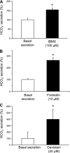
Effects of IBMX, forskolin, and genistein on esophageal HCO3 secretion. A: 3-isobutyl-1-methylxanthine (IBMX), a phosphodiesterase inhibitor, increased HCO3 secretion significantly (n = 6, P < 0.01). B: forskolin, an adenylyl cyclase activator, more than doubled HCO3 secretion (n = 6, P < 0.001). C: genistein an isoflavone phytoestrogen and a potentiator of CFTR increased HCO3 secretion significantly (n = 7, P < 0.005). *Significance compared with basal secretion.
Fig. 7.
Fig. 7.
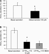
Effects of glibenclamide and CFTRinh-172 on esophageal HCO3 secretion. A: glibenclamide, a Cl channel (CFTR) blocker, inhibited basal HCO3 secretion significantly (n = 6, P < 0.05). B: CFTRinh-172 inhibited basal HCO3 secretion significantly and inhibited stimulation of secretion by IBMX. *Significance compared with basal secretion.
Fig. 8.
Fig. 8.
Immunofluorescence localization and RT-PCR amplification of the luminal anion exchanger SLC26A6 in SMG. A: staining of SLC26A6 (red) was positive at the luminal membrane. B: nuclei were counterstained with DAPI. C: merged image showing expression of the anion exchanger at the apical membrane of an interlobular duct. D: SLC26A6 expression was confirmed by RT-PCR using mRNA isolated from dissected SMG (lane 1) and trachea (lane 2) as positive control. The 494-bp band was the predicted product size.
Fig. 9.
Fig. 9.
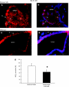
Immunofluorescence localization of Na+-K+-2Cl in the SMG using a mouse monoclonal antibody. In mucous cells, A shows intense staining (red) for Na+-K+-2Cl and B is a merged image of an acinus showing Na+-K+-2Cl and nuclei, counterstained blue with DAPI. These images indicate that Na+-K+-2Cl clearly delineated the basolateral membrane. In duct cells, C shows the localization of Na+-K+-2Cl (red) in interlobular duct cells of SMG, and D is the merged image of a duct showing Na+-K+-2Cl and nuclei stained blue with DAPI. The images indicate that Na+-K+-2Cl staining (red) was intense and clearly delineated the basolateral membrane. Bar = 5 μm. E: bumetanide, an Na+-K+-2Clblocker, decreased HCO3 secretion significantly (n = 3, P < 0.005). *Significance compared with basal secretion.
Fig. 10.
Fig. 10.
Cell model of a duct cell depicting ion transport mechanisms involved in HCO3 secretion in esophageal SMG. Basolateral HCO3 entry is mediated by Na+-(HCO3)n cotransporter (NBCe1). The anion exchanger Cl-HCO3 (AE2) mediates Cl uptake by the cell and could play a role in intracellular pH regulation. Basolateral Na+-K+-2Cl (NKCC1) mediates Na+, K+, and Cl entry into the cell. HCO3 efflux at the apical membrane is mediated by SLC26A6. Apical CFTR may serve as a channel that is permeable to HCO3 and therefore can contribute directly to HCO3 secretion. Alternatively, CFTR may serve predominantly as a channel for apical Cl efflux, a fraction of which may be shunted to drive apical Cl-HCO3 exchanger (SLC26A6) leading to HCO3 secretion. This model is based on data from this study and a previous study from our lab (2) where the role of different transport inhibitors, including carbonic anhydrase inhibitors on HCO3 secretion was evaluated.

References

    1. Abdulnour-Nakhoul S, Nakhoul NL, Orlando RC. Lumen-to-surface pH gradients in opossum and rabbit esophagi: role of submucosal glands. Am J Physiol Gastrointest Liver Physiol 278: G113–G120, 2000 - PubMed
    1. Abdulnour-Nakhoul S, Nakhoul NL, Wheeler SA, Wang P, Swenson ER, Orlando RC. HCO3− secretion in the esophageal submucosal glands. Am J Physiol Gastrointest Liver Physiol 288: G736–G744, 2005 - PubMed
    1. Abdulnour-Nakhoul S, Nakhoul NL, Wheeler SA, Haque S, Wang P, Brown K, Orlando GS, Orlando RC. Characterization of esophageal submucosal glands in pig tissue and cultures. Dig Dis Sci 52: 3054–3065, 2007 - PubMed
    1. Abuladze N, Lee I, Newman D, Hwang J, Boorer K, Pushkin A, Kurtz I. Molecular cloning, chromosomal localization, tissue distribution, and functional expression of the human pancreatic sodium bicarbonate cotransporter. J Biol Chem 273: 17689–17695, 1998 - PubMed
    1. Al Yassin TM, Toner PG. Fine structure of squamous epithelium and submucosal glands of human oesophagus. J Anat 123: 705–721, 1977 - PMC - PubMed

Publication types

MeSH terms

Substances

LinkOut - more resources