. 2011 May 27;286(21):18994-9000.
doi: 10.1074/jbc.M110.192286. Epub 2011 Apr 7.

A single amino acid residue constitutes the third dimerization domain essential for the assembly and function of the tetrameric polycystin-2 (TRPP2) channel

Affiliations

A single amino acid residue constitutes the third dimerization domain essential for the assembly and function of the tetrameric polycystin-2 (TRPP2) channel

Shuang Feng et al. J Biol Chem. .

Abstract

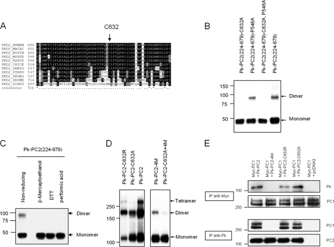
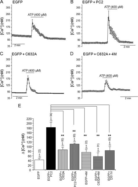
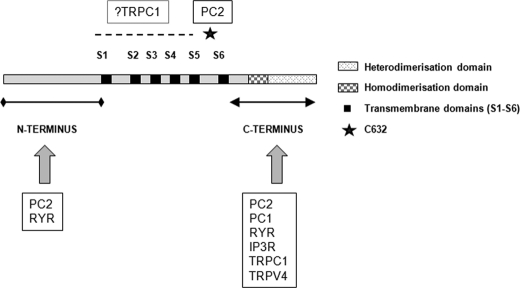
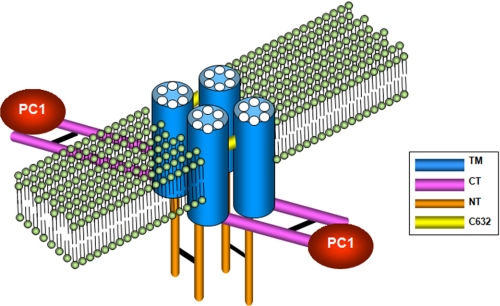
Autosomal dominant polycystic kidney disease (ADPKD), the most common inherited cause of kidney failure, is caused by mutations in either PKD1 (85%) or PKD2 (15%). The PKD2 protein, polycystin-2 (PC2 or TRPP2), is a member of the transient receptor potential (TRP) superfamily and functions as a nonselective calcium channel. PC2 has been found to form oligomers in native tissues, suggesting that similar to other TRP channels, it may form functional homo- or heterotetramers with other TRP subunits. We have recently demonstrated that the homodimerization of PC2 is mediated by both N-terminal and C-terminal domains, and it is known that PC2 can heterodimerize with PC1, TRPC1, and TRPV4. In this paper, we report that a single cysteine residue, Cys(632), mutated in a known PKD2 pedigree, constitutes the third dimerization domain for PC2. PC2 truncation mutants lacking both N and C termini could still dimerize under nonreducing conditions. Mutation of Cys(632) alone abolished dimerization in these mutants, indicating that it was the critical residue mediating disulfide bond formation between PC2 monomers. Co-expression of C632A PC2 mutants with wild-type PC2 channels reduced ATP-sensitive endoplasmic reticulum Ca(2+) release in HEK293 cells. The combination of C632A and mutations disrupting the C-terminal coiled-coil domain (Val(846), Ile(853), Ile(860), Leu(867) or 4M) nearly abolished dimer formation and ATP-dependent Ca(2+) release. However, unlike the 4M PC2 mutant, a C632A mutant could still heterodimerize with polycystin-1 (PC1). Our results indicate that PC2 homodimerization is regulated by three distinct domains and that these events regulate formation of the tetrameric PC2 channel.


Figures

FIGURE 1.
FIGURE 1.
Evidence of a third dimerization domain for PC2. A, expression pattern of epitope-tagged mutant PC2 on nonreducing SDS-PAGE. The truncation mutant PC2(224–968), which lacks the N terminus (1–223), is still able to form homodimers and tetramers. Mutation of CC2 (4M) in this construct results in a predominant monomeric pattern with faint but detectable dimers. Calnexin was used as an endogenous control for loading. B, deletion of the N terminus and first extracellular loop (469–968) shows a migration pattern similar to that of PC2(224–968) under nonreducing conditions. Mutation of CC2 (4M) results in a predominant monomeric pattern with faint dimers. C, diagram of PC2 showing its likely topology and key residues. The N-terminal dimerization domain (NTDD), coiled coil domains 1 (CC1) and 2 (CC2) within the C terminus have been described previously.
FIGURE 2.
FIGURE 2.
A conserved cysteine residue in the third extracellular loop mediates PC2 dimerization. A, sequence alignment of Cys632 showing its complete evolutionary conservation from human to Caenorhabditis elegans PC2. B, expression pattern of epitope-tagged mutant PC2 on nonreducing SDS-PAGE. Deletion of both N- and C-terminal domains, PC2(224–679), does not abolish dimer formation. Mutation at Cys632 but not Pro546 abolishes the formation of dimer formation in the absence of the two known dimerization domains. C, cell lysates of PC2(224–679) resolved on SDS-PAGE. Lysates were either incubated under nonreducing conditions, reducing agents (β-mercaptoethanol, DTT), or performic acid prior to loading. Immunoblotting was with anti-Pk tag antibody. D, expression pattern of epitope-tagged full-length PC2 on nonreducing SDS-PAGE. Wild-type PC2 migrates most prominently with dimeric and tetrameric species. The C632A mutation abolished tetramer bands but with an equal ratio of monomers and dimers. The double mutation (C632A+4M) almost completely abolished dimer formation. The pattern observed with C632R was intermediate between wild type and C632A. E, immunoprecipitation assays with epitope-tagged full-length PC1 and PC2. Mutation at C632A or C632R did not affect the PC1-PC2 interaction, whereas CC2 mutations (4M) abolished PC1-PC2 interactions.
FIGURE 3.
FIGURE 3.
The Cys632 mutation abrogates ATP-dependent PC2 Ca2+ release channel activity. A–D, averaged calcium transients in response to ATP application (400 μm) in HEK293T cells expressing EGFP (A), EGFP + PC2 (B), EGFP + PC2-C632A (C), and EGFP + PC2-C632A+4M (D). The gray horizontal bars indicate duration of ATP application. E, histogram showing the average rise in [Ca2+]i induced by ATP in cells expressing EGFP (0.5 μg of cDNA), EGFP + PC2 (0.5 μg of cDNA), EGFP + PC2-C632A, EGFP + PC2 + PC2-C632A, EGFP + PC2–4M, EGFP + PC2-C632A+4 M and EGFP + PC2-D511V. Bars represent mean ± S.E. *, p < 0.05; **, p < 0.01.
FIGURE 4.
FIGURE 4.
Diagrammatic representation of PC2 homophilic and heterophilic interaction domains. The various interaction domains between PC2 and a number of key calcium channel proteins are shown. Apart from PC2, PC1, and inositol trisphosphate receptor (IP3R), the key residues mediating other interactions have not been fully defined. RYR, ryanodine receptor.
FIGURE 5.
FIGURE 5.
Model of PC2 in a lipid bilayer showing the three dimerization domains. PC2 is displayed as a homotetramer with the transmembrane domains (TM) embedded in a lipid bilayer. C-terminal (CT) homodimerization mediated by the C-terminal CC2 (Ser835-Ala873) is indicated by a black line. CC2-mediated dimerization has a specific role in PC1 recognition and formation of a PC1-PC2 heteromeric complex. N-terminal (NT) dimerization mediated by the N-terminal dimerization domain (Gly199-Glu207) is indicated by a black line. Disulfide bonding between PC2 monomers mediated by Cys632 is indicated by a yellow line.

References

    1. Calvet J. P., Grantham J. J. (2001) Semin. Nephrol. 21, 107–123 - PubMed
    1. Wilson P. D. (2004) N. Engl. J. Med. 350, 151–164 - PubMed
    1. Mochizuki T., Wu G., Hayashi T., Xenophontos S. L., Veldhuisen B., Saris J. J., Reynolds D. M., Cai Y., Gabow P. A., Pierides A., Kimberling W. J., Breuning M. H., Deltas C. C., Peters D. J., Somlo S. (1996) Science 272, 1339–1342 - PubMed
    1. Montell C., Birnbaumer L., Flockerzi V., Bindels R. J., Bruford E. A., Caterina M. J., Clapham D. E., Harteneck C., Heller S., Julius D., Kojima I., Mori Y., Penner R., Prawitt D., Scharenberg A. M., Schultz G., Shimizu N., Zhu M. X. (2002) Mol. Cell 9, 229–231 - PubMed
    1. Tsiokas L., Arnould T., Zhu C., Kim E., Walz G., Sukhatme V. P. (1999) Proc. Natl. Acad. Sci. U.S.A. 96, 3934–3939 - PMC - PubMed

Publication types

MeSH terms