Comparative Study
. 2011 Jul;338(1):82-91.
doi: 10.1124/jpet.111.180620. Epub 2011 Apr 7.

Catalase and superoxide dismutase conjugated with platelet-endothelial cell adhesion molecule antibody distinctly alleviate abnormal endothelial permeability caused by exogenous reactive oxygen species and vascular endothelial growth factor

Affiliations
Comparative Study

Catalase and superoxide dismutase conjugated with platelet-endothelial cell adhesion molecule antibody distinctly alleviate abnormal endothelial permeability caused by exogenous reactive oxygen species and vascular endothelial growth factor

Jingyan Han et al. J Pharmacol Exp Ther. 2011 Jul.

Abstract

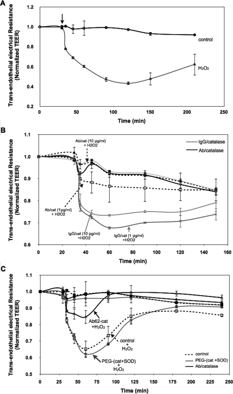
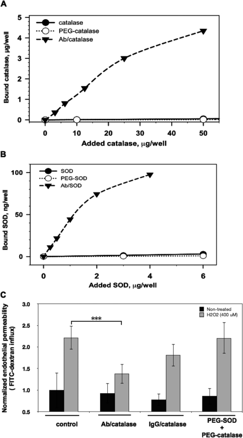
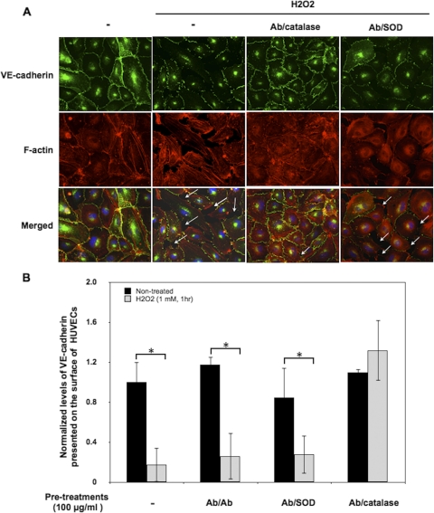
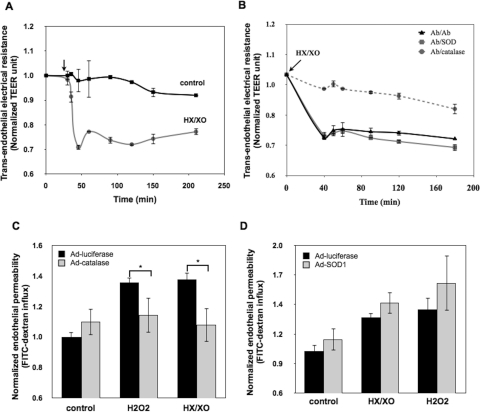
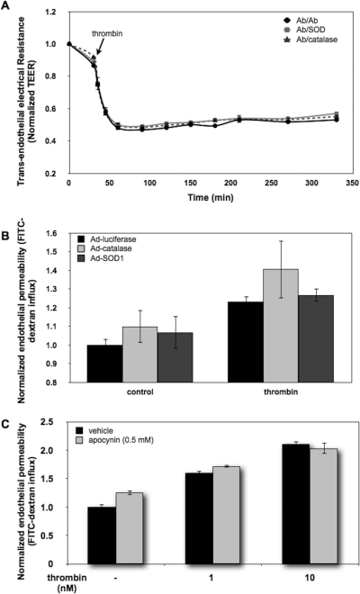
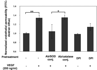
Reactive oxygen species (ROS) superoxide anion (O(2)()) and hydrogen peroxide (H(2)O(2)) produced by activated leukocytes and endothelial cells in sites of inflammation or ischemia cause endothelial barrier dysfunction that may lead to tissue edema. Antioxidant enzymes (AOEs) catalase and superoxide dismutase (SOD) conjugated with antibodies to platelet-endothelial cell adhesion molecule-1 (PECAM-1) specifically bind to endothelium, quench the corresponding ROS, and alleviate vascular oxidative stress and inflammation. In the present work, we studied the effects of anti-PECAM/catalase and anti-PECAM/SOD conjugates on the abnormal permeability manifested by transendothelial electrical resistance decline, increased fluorescein isothiocyanate-dextran influx, and redistribution of vascular endothelial-cadherin in human umbilical vein endothelial cell (HUVEC) monolayers. Anti-PECAM/catalase protected HUVEC monolayers against H(2)O(2)-induced endothelial barrier dysfunction. Polyethylene glycol-conjugated catalase exerted orders of magnitude lower endothelial uptake and no protective effect, similarly to IgG/catalase. Anti-PECAM/catalase, but not anti-PECAM/SOD, alleviated endothelial hyperpermeability caused by exposure to hypoxanthine/xanthine oxidase, implicating primarily H(2)O(2) in the disruption of the endothelial barrier in this model. Thrombin-induced endothelial permeability was not affected by treatment with anti-PECAM/AOEs or the NADPH oxidase inhibitor apocynin or overexpression of AOEs, indicating that the endogenous ROS play no key role in thrombin-mediated endothelial barrier dysfunction. In contrast, anti-PECAM/SOD, but not anti-PECAM/catalase, inhibited a vascular endothelial growth factor (VEGF)-induced increase in endothelial permeability, identifying a key role of endogenous O(2)() in the VEGF-mediated regulation of endothelial barrier function. Therefore, AOEs targeted to endothelial cells provide versatile molecular tools for testing the roles of specific ROS in vascular pathology and may be translated into remedies for these ROS-induced abnormalities.


Figures

Fig. 1.
Fig. 1.
Anti-PECAM/catalase conjugates protect against endothelial barrier dysfunction induced by H2O2 insult. A, TEER across HUVEC monolayers was used to evaluate endothelial permeability. HUVEC monolayers grown onto Transwell inserts (3.0-μm pore size) were transferred to Endohm (World Precision Instruments) chambers for resistance measurement at different time points. At the arrow-indicated time point, the cells were stimulated with H2O2 (400 μM). The recorded values were normalized to the initial resistances of the monolayers before H2O2 treatment. The normalized TEER values are represented as means ± S.D. (n = 3). B, Ab/catalase treatment (1 and 10 μg/ml) abolished H2O2-induced endothelial hyperpermeability. HUVEC monolayers were preincubated with conjugates for 30 min and washed three times to remove unbound conjugates. Cells then were stimulated with H2O2 (400 μM) and subjected to TEER measurement. C, mixture of PEGylated SOD and catalase (10 μg/ml of each) failed to attenuate H2O2-induced TEER decline.
Fig. 2.
Fig. 2.
PEGylated SOD and catalase do not bind to endothelial cells and do not inhibit H2O2-stimulated endothelial permeability for macromolecules. A, Ab/catalase, but not PEGylated catalase, specifically bound to HUVECs. B, Ab/SOD, but not PEGylated SOD, specifically targeted HUVECs. C, Ab/catalase, but not PEGylated SOD and catalase, alleviated H2O2-induced endothelial permeability for macromolecules as measured by the FITC-dextran influx assay. Values are the normalized percentages of total FITC-dextran passing across cells relative that of the control group. Error bars represent the S.D. (n = 3; ***, p < 0.001).
Fig. 3.
Fig. 3.
Ab/catalase inhibits H2O2-induced redistribution of VE-cadherin. A, immunocytochemical staining of VE-cadherin and F-actin in HUVECs. Confluent HUVECs treated with Ab/Ab, Ab/catalase, and Ab/SOD conjugate (100 μg/ml, 30 min) were washed and stimulated with H2O2 (1 mM) for 1 h. Cells then were washed, fixed, and immunostained for VE-cadherin (green fluorescence) and F-actin (red fluorescence). Arrows point to the intercellular gaps. B, whole-cell ELISA was used to quantify VE-cadherin presented on the cell surface. The data are normalized to control cells without H2O2 stimulation and are means from three experiments ± S.D. (*, p < 0.05 in a Student's t test).
Fig. 4.
Fig. 4.
Ab/catalase, but not Ab/SOD, specifically inhibited superoxide-induced endothelial permeability. A, superoxide anion generated by the HX/XO system increased endothelial permeability. HUVEC monolayers were exposed to HX (200 μM) and XO (20 mU), and the TEER values at indicated time points were recorded. The data shown are normalized TEER values relative to the initial TEER values of the monolayers before stimulation. Means ± S.D. are shown. (n = 3). B, Ab/catalase, but not Ab/SOD, rescued superoxide-induced endothelial permeability. HUVECs were treated with drug-free conjugates Ab/Ab (100 μg/ml), Ab/catalase (10 μg/ml), and Ab/SOD (100 μg/ml). Cells then were washed and stimulated with HX/XO. Only Ab/catalase treatment reversed HX/XO-induced TEER decline. C, overexpression of cytosolic catalase significantly attenuated endothelial permeability induced by HX/XO. HUVECs overexpressing luciferase (negative control) or catalase were stimulated with H2O2 (600 μM) or HX/XO (400 μM/40 mU) for 90 min followed by FITC-dextran influx measurement. Data shown are normalized dextran influx (fold of basal) and are means ± S.D. (n = 6; *, p < 0.05). D, overexpression of cytosolic SOD1 did not show protective effects on superoxide anion- or H2O2-induced hyperpermeability. HUVECs overexpressing SOD1 were treated with HX/XO (400 μM/40 mU) or H2O2 (600 μM) for 90 min. The endothelial permeability to FITC-dextran was measured.
Fig. 5.
Fig. 5.
Thrombin-induced intracellular ROS is not involved in the regulation of endothelial permeability. A, pretreatment of Ab/catalase and Ab/SOD (100 μg/ml, 30 min) did not attenuate thrombin-induced endothelial permeability. Conjugate-treated HUVEC monolayers were exposed to thrombin (20 nM), and the TEER values of the monolayers were measured at the indicated time points. Data shown are means ± S.D. (n = 3). B, overexpression of cytosolic catalase and SOD1 did not modulate thrombin-induced endothelial permeability. HUVECs expressing cytosolic luciferase serving as a control, catalase, and SOD1 by adenoviral infection were stimulated with thrombin (40 nM, 1 h) followed by the FITC-dextran influx assay. C, blockage of thrombin-induced ROS generation did not affect endothelial permeability. HUVEC monolayers were preincubated with dimethyl sulfoxide (vehicle) or apocynin (0.5 mM) for 1 h and then exposed to thrombin (1 and 10 nM) for 1 h. Endothelial permeability to macromolecules was assessed by the FITC-dextran influx assay. Thrombin stimulation increased permeability in a dose-dependent manner, and no significant difference was detected between control and apocynin-treated cells.
Fig. 6.
Fig. 6.
Ab/SOD conjugate specifically protects the endothelial barrier from VEGF insult. Ab/SOD, but not Ab/catalase, inhibited VEGF-induced endothelial permeability. HUVEC monolayers were preincubated with conjugates of Ab/catalase and Ab/SOD (100 μg/ml, 30 min) or DPI (10 μM, 1 h). Cells then were stimulated with VEGF (200 ng/ml) for 30 min followed by the FITC-dextran influx assay. Data shown are means ± S.D. (n = 4; *, p < 0.05; **, p < 0.01).

References

    1. Alexander JS, Alexander BC, Eppihimer LA, Goodyear N, Haque R, Davis CP, Kalogeris TJ, Carden DL, Zhu YN, Kevil CG. (2000) Inflammatory mediators induce sequestration of VE-cadherin in cultured human endothelial cells. Inflammation 24:99–113 - PubMed
    1. Alom-Ruiz SP, Anilkumar N, Shah AM. (2008) Reactive oxygen species and endothelial activation. Antioxid Redox Signal 10:1089–1100 - PubMed
    1. Atochina EN, Balyasnikova IV, Danilov SM, Granger DN, Fisher AB, Muzykantov VR. (1998) Immunotargeting of catalase to ACE or ICAM-1 protects perfused rat lungs against oxidative stress. Am J Physiol 275:L806–L817 - PubMed
    1. Bae JS, Yang L, Manithody C, Rezaie AR. (2007) Engineering a disulfide bond to stabilize the calcium-binding loop of activated protein C eliminates its anticoagulant but not its protective signaling properties. J Biol Chem 282:9251–9259 - PubMed
    1. Barnard ML, Matalon S. (1992) Mechanisms of extracellular reactive oxygen species injury to the pulmonary microvasculature. J Appl Physiol 72:1724–1729 - PubMed

Publication types

MeSH terms

LinkOut - more resources