

Value of MR venography for detection of internal jugular vein anomalies in multiple sclerosis: a pilot longitudinal study
- PMID: 21474626
- PMCID: PMC7965543
- DOI: 10.3174/ajnr.A2386
Value of MR venography for detection of internal jugular vein anomalies in multiple sclerosis: a pilot longitudinal study
Abstract
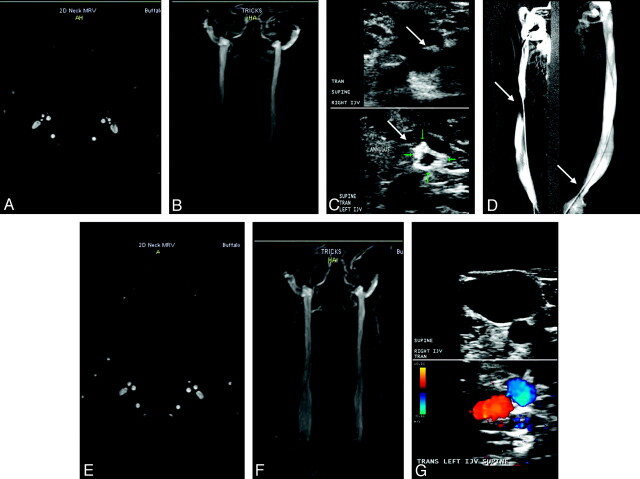
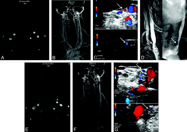
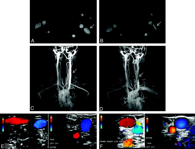
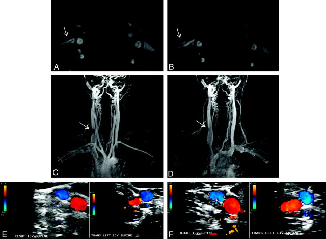
Background and purpose: CCSVI was recently described in patients with MS. CCSVI is diagnosed noninvasively by Doppler sonography and invasively by catheter venography. We assessed the role of conventional MRV for the detection of IJV anomalies in patients with MS diagnosed with CCSVI and in healthy controls who underwent MRV and Doppler sonography examinations during 6 months.
Materials and methods: Ten patients with MS underwent TOF, TRICKS, Doppler sonography, and catheter venography at baseline. They were treated at baseline with percutaneous angioplasty and re-evaluated 6 months' posttreatment with MRV and Doppler sonography. In addition, 6 healthy controls underwent a baseline and a 6-month follow-up evaluation by Doppler sonography and MRV.
Results: At baseline, the sensitivity, specificity, PPV, and NPV of Doppler sonography for detecting IJV abnormalities relative to catheter venography in patients with MS were calculated, respectively, at 82%, 100%, 99%, and 95%. The figures were 99%, 33%, 33%, 99% for TOF and 99%, 39%, 35%, and 99% for TRICKS. Venous anomalies included the annulus, septum, membrane, and malformed valve. No agreement was found between TOF and catheter venography in 70% of patients with MS and between TRICKS and catheter venography in 60% of patients with MS. At follow-up, 50% of the patients with MS presented with abnormalities on Doppler sonography but only 30% were diagnosed with restenosis.
Conclusions: Conventional MRV has limited value for assessing IJV anomalies for both diagnostic and posttreatment purposes.
Figures
References
-
- Zamboni P, Menegatti E, Galeotti R, et al. The value of cerebral Doppler venous haemodynamics in the assessment of multiple sclerosis. J Neurol Sci 2009;282:21–27. Epub 2009 Jan 13 - PubMed
-
- Zamboni P, Galeotti R, Menegatti E, et al. A prospective open-label study of endovascular treatment of chronic cerebrospinal venous insufficiency. J Vasc Surg 2009;50:1348–58 e1–3 - PubMed
-
- Lee AB, Laredo J, Neville R. Embryological background of truncular venous malformation in the extracranial venous pathways as the cause of chronic cerebro spinal venous insufficiency. Int Angiol 2010;29:95–108 - PubMed
-
- Al-Omari MH, Rousan LA. Internal jugular vein morphology and hemodynamics in patients with multiple sclerosis. Int Angiol 2010;29:115–20 - PubMed
Publication types
MeSH terms
LinkOut - more resources
Full Text Sources
Medical