. 2011 Jul 1;184(1):37-49.
doi: 10.1164/rccm.201010-1637OC. Epub 2011 Apr 7.

Cyclooxygenase-2 regulates Th17 cell differentiation during allergic lung inflammation

Affiliations

Cyclooxygenase-2 regulates Th17 cell differentiation during allergic lung inflammation

Hong Li et al. Am J Respir Crit Care Med. .

Abstract

Rationale: Th17 cells comprise a distinct lineage of proinflammatory T helper cells that are major contributors to allergic responses. It is unknown whether cyclooxygenase (COX)-derived eicosanoids regulate Th17 cells during allergic lung inflammation.

Objectives: To determine the role of COX metabolites in regulating Th17 cell differentiation and function during allergic lung inflammation.

Methods: COX-1(-/-), COX-2(-/-), and wild-type mice were studied in an in vivo model of ovalbumin-induced allergic inflammation and an in vitro model of Th17 differentiation using flow cytometry, cytokine assays, confocal microscopy, real-time polymerase chain reaction, and immunoblotting. In addition, the role of specific eicosanoids and their receptors was examined using synthetic prostaglandins (PGs), selective inhibitors, and siRNA knockdown.

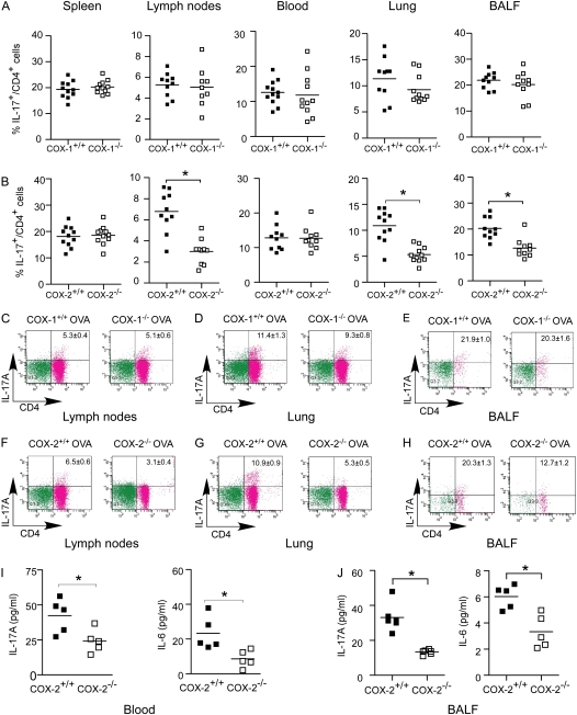
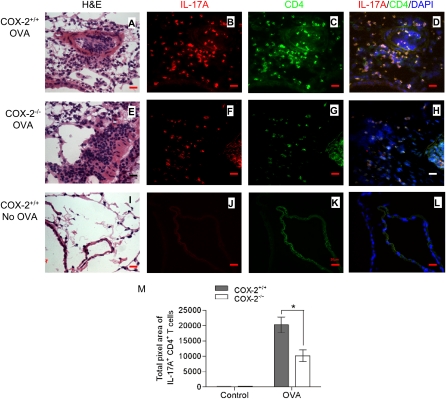
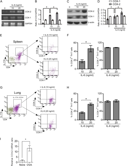
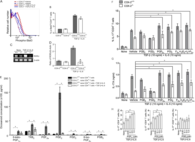
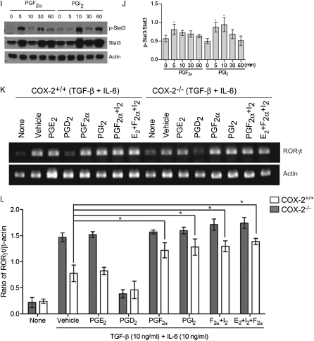
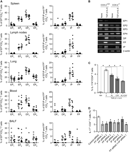
Measurements and main results: Th17 cell differentiation in lung, lymph nodes, and bronchoalveolar lavage fluid was significantly lower in COX-2(-/-) mice after ovalbumin sensitization and exposure in vivo. In vitro studies revealed significantly impaired Th17 cell differentiation of COX-2(-/-) naive CD4(+) T cells with decreased Stat3 phosphorylation and RORγt expression. Synthetic PGF(2α) and PGI(2) enhanced Th17 cell differentiation of COX-2(-/-) CD4(+) T cells in vitro. The selective COX-2 inhibitor, NS-398, and PGF(2α) receptor and PGI(2) receptor siRNA knockdown significantly decreased Th17 cell differentiation in vitro. Administration of synthetic PGs restored accumulation of Th17 cells in lungs of allergic COX-2(-/-) mice in vivo.

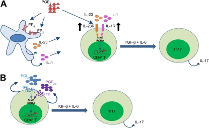
Conclusions: COX-2 is a critical regulator of Th17 cell differentiation during allergic lung inflammation via autocrine signaling of PGI(2) and PGF(2α) through their respective cell surface receptors.


Figures

Figure 1.
Figure 1.
Reduced Th17 cells in lung, bronchoalveolar lavage fluid (BALF), and lymph nodes of cyclooxygenase (COX)-2−/− mice after ovalbumin (OVA) sensitization and exposure in vivo. COX-1+/+, COX-1−/−, COX-2+/+, and COX-2−/− mice (n = 9–12 each) were sensitized with OVA in adjuvant. Fourteen to 21 days later, mice were exposed to inhaled OVA for 4 consecutive days. The percentages of IL-17A+ CD4+ T cells in spleen, lymph nodes, blood, lung, and BALF from COX-1+/+ versus COX-1−/− mice (A) and COX-2+/+ versus COX-2−/− mice (B) were analyzed by flow cytometry 48 hours after the last OVA exposure. Flow cytometry scattergrams show that the percentage of IL-17A+ CD4+ cells were similar in COX-1+/+ versus COX-1−/− lymph nodes (C), lung (D), and BALF (E). In contrast, COX-2−/− mice had significantly fewer IL-17A+ CD4+ cells in lymph nodes (F), lung (G), and BALF (H). IL-17A and IL-6 concentrations in blood (I) and BALF (J) were measured by ELISA and BioPlex assay 48 hours after the last OVA exposure. For panels A, B, and I, lines indicate the mean, and each symbol (solid squares, COX-1+/+ or COX-2+/+; open squares, COX-1−/− or COX-2−/−) represents an individual mouse. *P < 0.05 versus wild-type.
Figure 2.
Figure 2.
Reduced Th17 cells in lung of cyclooxygenase (COX)-2−/− mice after ovalbumin (OVA) exposure. COX-2+/+ and COX-2−/− mice were sensitized with OVA in adjuvant (or given adjuvant alone). Fourteen to 21 days later, mice were exposed to inhaled OVA (or inhaled phosphate-buffered saline) for 4 consecutive days. Forty-eight hours after final OVA (or phosphate-buffered saline) exposure, lung tissue sections were stained with hematoxylin and eosin (H&E) and visualized by light microscopy (A, E, and I) (original magnification ×100). Visualization of Th17 cells in COX-2+/+ and COX-2−/− lung tissue sections was accomplished by immunofluorescence staining with phycoerythrin (PE)-labeled anti–IL-17A (B, F, and J) and fluorescein isothiocyanate (FITC)–labeled anti-CD4 (C, G, and K) antibodies. D, H, and L are merged images of anti–IL-17A and PE, anti–CD4 and FITC, and DAPI. All immunofluorescent images are shown at original magnification ×60; numerical aperture, 1.4; scale bar, 25 μm. Results are representative of three independent experiments. Multiple (n = 22–29) randomly selected regions (1,300 × 1,030 pixel2 = 350 × 277 μm2) from each lung section were counted and quantified by a masked observer using MetaMorph software (M); *P < 0.05 versus COX-2+/+.
Figure 3.
Figure 3.
Regulation of cyclooxygenase (COX)-1 and COX-2 during Th17 cell differentiation from naive CD4+ T cells. Naive CD4+ T cells isolated from spleen were stimulated with or without anti-CD3 (3 μg/ml), anti-CD28 (3 μg/ml), anti–INF-γ (3 μg/ml), transforming growth factor-β (10 ng/ml), and IL-6 (10 or 20 ng/ml) for 4 days to induced Th17 differentiation and then the cells were analyzed for COX-1 and COX-2 expression by real-time polymerase chain reaction (RT-PCR), protein immunoblotting, and flow cytometry. (A) PCR products showing mRNA levels of COX-1, COX-2, and glyceralhehyde phosphate dehydrogenase (GAPDH). (B) COX-1 and COX-2 mRNAs were quantified by densitometry and expressed as the density ratio of COX-1 or COX-2 relative to GAPDH. Results are representative of three independent experiments. (C) Immunoblot analysis of cell lysates for COX-1, COX-2, and β-actin. (D) Protein levels of COX-1 and COX-2 were quantified by densitometry and expressed relative to β-actin. Results are representative of three independent experiments. (E and G) Naive CD4+ T cells were stimulated with anti-CD3, anti-CD28, anti–INF-γ, transforming growth factor-β, and IL-6 (10 and 20 ng/ml) for 4 days, and then stimulated for 4 hours with 12–0-tetradecanoyl-phorbol-13-acetate and ionomycin (500 ng/ml each) in the presence of brefeldin A (1 μg/ml) before intracellular staining with anti-CD4, IL-17, COX-1, and COX-2. Gating on the CD4+ IL-17A+ cell population, COX-1 and COX-2 expression was analyzed by flow cytometry. (F and H) The percent of the COX-1– and COX-2–positive Th17 cells after treatment with different concentrations of IL-6 is shown. Results are representative of three independent experiments. *P < 0.05. (I) Lung CD4+ T cells were isolated after ovalbumin (OVA) exposure in vivo and COX-2 mRNA levels were quantified by RT-PCR; n = 6; *P < 0.01 versus control (no OVA).
Figure 4.
Figure 4.
Impaired Th17 differentiation in cyclooxygenase (COX)-2−/− naive CD4+ T cells in vitro. (A) Naive CD4+ T cells isolated from spleens of COX-2+/+ and COX-2−/− mice were treated with or without anti-CD3 (3 μg/ml), anti-CD28 (3 μg/ml), anti–INF-γ (3 μg/ml), transforming growth factor (TGF)-β (10 ng/ml), and IL-6 (10 ng/ml) for 4 days. The cells were then stimulated for 4 hours with 12–0-tetradecanoyl-phorbol-13-acetate and inomycin (500 ng/ml each) in the presence of brefeldin A (1 μg/ml) before flow cytometry analysis. Data are representative of three independent experiments. (B) The percentage of naive CD4+ T cells from COX-2+/+ and COX-2−/− mice that differentiated to CD4+ IL-17A+ (Th17) cells is shown. (C) Inhibition of COX-2 with NS-398 during Th17 cell differentiation from naive CD4+ T cells was examined. Naive CD4+ T cells were cultured with anti-CD3 (3 μg/ml), anti-CD28 (3 μg/ml), anti–INF-γ (3 μg/ml), TGF-β (10 ng/ml), and IL-6 (10 ng/ml) in the presence or absence of NS-398 (20 μM) for 4 days. The cells were then stimulated for 4 hours with 12–0-tetradecanoyl-phorbol-13-acetate and inomycin (500 ng/ml each) in the presence of brefeldin A (1 μg/ml) before flow cytometry analysis. (D) The percentage of IL-17A+ CD4+ cells was examined by flow cytometry at different time points (Days 1–7) after treatment with anti-CD3, anti-CD28, anti–INF-γ, TGF-β, and IL-6. (E) Cell culture supernatants from D were collected at different time points and IL-17A levels were assayed by ELISA. (F) Naive CD4+ T cells from COX-2+/+ and COX-2−/− mice were differentiated to Th17 cells in the presence of different amounts of IL-6 (0, 5, 10, and 20 ng/ml) for 4 days and the percent of CD4+ IL-17A+ cells was analyzed by flow cytometry. (G) IL-17A levels in cell culture supernatants from F were assayed by ELISA. For panels CF, data are representative of three independent experiments; closed circles = COX-2+/+; open circles = COX-2−/−; * P < 0.05 versus COX-2+/+. (H) Total number of CD4+ T cells were enumerated in spleen, lymph nodes, and lung from COX-2+/+ and COX-2−/− mice (n = 10 per group). (I) Splenocytes from COX-2+/+ and COX-2−/− mice were analyzed for CD62L+ and CD4+ expression. Representative scattergrams are illustrated and the percentages of CD62L+ CD4+ cells (mean ± SE; n = 10 per group) are shown. (J) Naive CD4+ CD62L+ T cells from spleens of COX-2+/+ and COX-2−/− mice were first sorted by flow cytometry. These highly purified naive CD4+ CD62L+ T cells were then differentiated into Th17 cells as described in A. On Day 4, the cells were collected and the percentage of IL-17A+ CD4+ cells was analyzed by flow cytometry. (K) Quantification of Th17 cell differentiation of COX-2+/+ and COX-2−/− naive CD4+ CD62L+ T cells is shown.
Figure 5.
Figure 5.
Cyclooxygenase (COX)-2–derived prostaglandins (PGs) regulate Th17 cell differentiation in vitro. (A) Stat3 phosphorylation during Th17 differentiation of CD4+ T cells from COX-2+/+ and COX-2−/− mice was assayed using anti–phospho-stat3 (pY705)-PE by flow cytometry. (B) Ratio of phospho-Stat3/total Stat3 during Th17 cell differentiation of COX-2\\plus/\plus+/+ and COX-2−/− naive CD4+ T cells. (C) RORγt mRNA expression in differentiated CD4+ T cells from COX-2+/+ and COX-2−/− mice was analyzed by real-time polymerase chain reaction. (D) Ratio of RORγt to β-actin mRNA during Th17 cell differentiation of COX-2+/+ and COX-2−/− naive CD4+ CD62L+T cells. All data are representative of three independent experiments; #P < 0.05 versus COX-2+/+ naive CD4+ T cells; *P < 0.05 versus COX-2+/+. (E) Eicosanoid levels in supernatants of naive CD4+ T cells and in vitro differentiated Th17 cells from COX-2+/+ and COX-2−/− mice were measured by liquid chromatography/tandem mass spectroscopy. Results are representative of five independent experiments. Effect of synthetic PGs on Th17 cell differentiation of (F) and IL-17A production by (G) naive CD4+ T cells isolated from COX-2+/+ and COX-2−/− mice in vitro. PGs were added at 1 μM on Days 2 and 3 of culture, and culture supernatants were collected on Day 4. IL-17A was measured by ELISA. Results are representative of three independent experiments; *P < 0.05 versus vehicle. (H) Dose dependency of PGI2, PGF, and PGE2 (50 nM to 1 μM) on Th17 cell differentiation was examined in vitro; n = 4; *P < 0.05 versus no PG. (I and J) Phospho-Stat3 and total Stat3 levels were determined by immunoblotting before and 5–60 minutes after treatment of naive CD4+ T cells with synthetic PGs (1 μM each). Results are representative of three independent experiments; * P < 0.05 versus time 0. (K and L) RORγt mRNA levels measured by real-time polymerase chain reaction in in vitro differentiated Th17 cells treated with or without synthetic PGs (1 μM each). Results are representative of three independent experiments; * P < 0.05 versus vehicle.
Figure 5.
Figure 5.
Cyclooxygenase (COX)-2–derived prostaglandins (PGs) regulate Th17 cell differentiation in vitro. (A) Stat3 phosphorylation during Th17 differentiation of CD4+ T cells from COX-2+/+ and COX-2−/− mice was assayed using anti–phospho-stat3 (pY705)-PE by flow cytometry. (B) Ratio of phospho-Stat3/total Stat3 during Th17 cell differentiation of COX-2\\plus/\plus+/+ and COX-2−/− naive CD4+ T cells. (C) RORγt mRNA expression in differentiated CD4+ T cells from COX-2+/+ and COX-2−/− mice was analyzed by real-time polymerase chain reaction. (D) Ratio of RORγt to β-actin mRNA during Th17 cell differentiation of COX-2+/+ and COX-2−/− naive CD4+ CD62L+T cells. All data are representative of three independent experiments; #P < 0.05 versus COX-2+/+ naive CD4+ T cells; *P < 0.05 versus COX-2+/+. (E) Eicosanoid levels in supernatants of naive CD4+ T cells and in vitro differentiated Th17 cells from COX-2+/+ and COX-2−/− mice were measured by liquid chromatography/tandem mass spectroscopy. Results are representative of five independent experiments. Effect of synthetic PGs on Th17 cell differentiation of (F) and IL-17A production by (G) naive CD4+ T cells isolated from COX-2+/+ and COX-2−/− mice in vitro. PGs were added at 1 μM on Days 2 and 3 of culture, and culture supernatants were collected on Day 4. IL-17A was measured by ELISA. Results are representative of three independent experiments; *P < 0.05 versus vehicle. (H) Dose dependency of PGI2, PGF, and PGE2 (50 nM to 1 μM) on Th17 cell differentiation was examined in vitro; n = 4; *P < 0.05 versus no PG. (I and J) Phospho-Stat3 and total Stat3 levels were determined by immunoblotting before and 5–60 minutes after treatment of naive CD4+ T cells with synthetic PGs (1 μM each). Results are representative of three independent experiments; * P < 0.05 versus time 0. (K and L) RORγt mRNA levels measured by real-time polymerase chain reaction in in vitro differentiated Th17 cells treated with or without synthetic PGs (1 μM each). Results are representative of three independent experiments; * P < 0.05 versus vehicle.
Figure 6.
Figure 6.
Administration of synthetic prostaglandins (PGs) restores Th17 cell percentages in allergic cyclooxygenase (COX)-2−/− mice in vivo. Alzet minipumps filled with PGE2, PGF, iloprost, or the combination of PGs were implanted into sensitized COX-2−/− mice. After 1 week, mice were exposed to ovalbumin daily for 4 days. Forty-eight hours after the last ovalbumin exposure, the mice were killed and the percentages of IL-17A+ CD4+ T cells in lung (A), bronchoalveolar lavage fluid (BALF) (B), and lymph nodes (C) were determined by flow cytometry. (D) IL-17 levels in BALF samples from the above mice were assayed for IL-17A by ELISA. Results are representative of three independent experiments. n = 4; *P < 0.05 versus saline vehicle. In A, B, and C, lines indicate the mean, and each symbol represents an individual mouse.
Figure 7.
Figure 7.
Expression of prostaglandin (PG) receptors on CD4+ T cells in vivo and in vitro differentiated Th17 cells from cyclooxygenase (COX)-2+/+ and COX-2−/− mice. (A) The percentage of CD4+ T cells isolated from spleen, lymph nodes, lung, blood, and bronchoalveolar lavage fluid (BALF) of allergic COX-2+/+ and COX-2−/− mice was determined by flow cytometry. Antibodies to each of the PG receptors were conjugated with AlexaFlur-488 or AlexaFlur-594 using a Therm Scientific DyLightTM 488/594 microscale antibody labeling kit. Cell suspensions were prepared and stained with the conjugated receptor antibodies. EP1-EP4, DP1, DP2, FP, and IP receptor-positive CD4+ T cells were quantified by flow cytometry by gating on the IL-17+ CD4+ T cell population. Lines indicate the mean, and each symbol (open squares, COX-2+/+; solid squares, COX-2−/−) represents an individual mouse. (B) Levels of EP1-EP4, DP, FP, and IP receptor mRNAs in naive CD4+ T cells and in in vitro differentiated Th17 cells from COX-2+/+ and COX-2−/− mice. Naive CD4+ T cells isolated from COX-2+/+ and COX-2−/− mice were stimulated with or without anti-CD3, anti-CD28, anti–INF-γ, IL-6, and transforming growth factor (TGF)-β for 4 days. Total RNA was then extracted and reverse transcribed to cDNA for detection of EP1-EP4, DP, FP, and IP receptor expression by real-time polymerase chain reaction. (C) The effects of FP and IP receptor antagonists on Th17 cell differentiation of naive CD4+ T cells were investigated. Naive CD4+ T cells from wild-type mice were differentiated to Th17 cells in the presence or absence of the FP receptor antagonist, AL8810, the IP receptor antagonist, CAY10441, or a combination of AL8810 and CAY10441. The percentage of Th17 cells was analyzed by flow cytometry after 5 days. n = 3; * P < 0.05. (D) The effects of FP and IP receptor knockdown on Th17 differentiation of naive CD4+ T cells were investigated. Naive CD4+ T cells from wild-type mice were transfected with IP receptor siRNAs, FP receptor siRNAs, a combination of IP receptor and FP receptor siRNAs, or control siRNA. Transfected cells were then differentiated in the presence of anti-CD3 (3 μg/ml), anti-CD28 (3 μg/ml), anti–INF-γ (3 μg/ml), TGF-β (10 ng/ml), and IL-6 (10 ng/ml) for 5 days and the percentage of Th17 cells was analyzed by flow cytometry. n = 3; * P < 0.05 versus control siRNA.
Figure 8.
Figure 8.
Proposed mechanisms for the regulation of Th17 cell differentiation by cyclooxygenase (COX)-2–derived prostaglandins (PGs). (A) Paracrine pathway of PGE2 effects on Th17 cell differentiation. Binding of PGE2 to EP2 and EP4 on dendritic cells leads to increased IL-23 and IL-1 expression. In addition, PGE2 induces IL-23R and IL-1R expression on CD4+ T cells. The IL-23 and IL-1 signals lead to activation of Stat3 and up-regulation of RORγt, which in conjunction with transforming growth factor (TGF)-β and IL-6, leads to IL-17 expression. (B) Autocrine pathway of PGI2 and PGF effects on Th17 cell differentiation. Binding of PGI2 and PGF to IP and FP receptors on CD4+ T cells leads to activation of Stat3 and up-regulation of RORγt, which in conjunction with TGF-β and IL-6 leads to IL-17 expression.

Comment in

References

    1. Adamson AS, Collins K, Laurence A, O'Shea JJ. The current status of lymphocyte signaling: new roles for old players. Curr Opin Immunol 2009;21:161–166 - PMC - PubMed
    1. Annunziato F, Cosmi L, Santarlasci V, Maggi L, Liotta F, Mazzinghi B, Parente E, Fili L, Ferri S, Frosali F, et al. Phenotypic and functional features of human Th17 cells. J Exp Med 2007;204:1849–1861 - PMC - PubMed
    1. Annunziato F, Cosmi L, Liotta F, Maggi E, Romagnani S. Human Th17 cells: are they different from murine Th17 cells? Eur J Immunol 2009;39:637–640 - PubMed
    1. Manel N, Unutmaz D, Littman DR. The differentiation of human Th17 cells requires transforming growth factor-beta and induction of the nuclear receptor RORγt. Nat Immunol 2008;9:641–649 - PMC - PubMed
    1. Iwakura Y, Nakae S, Saijo S, Ishigame H. The roles of IL-17A in inflammatory immune responses and host defense against pathogens. Immunol Rev 2008;226:57–79 - PubMed

Publication types

MeSH terms