

Activated protein C up-regulates procoagulant tissue factor activity on endothelial cells by shedding the TFPI Kunitz 1 domain
- PMID: 21474669
- PMCID: PMC3122952
- DOI: 10.1182/blood-2010-10-316257
Activated protein C up-regulates procoagulant tissue factor activity on endothelial cells by shedding the TFPI Kunitz 1 domain
Abstract
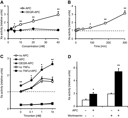
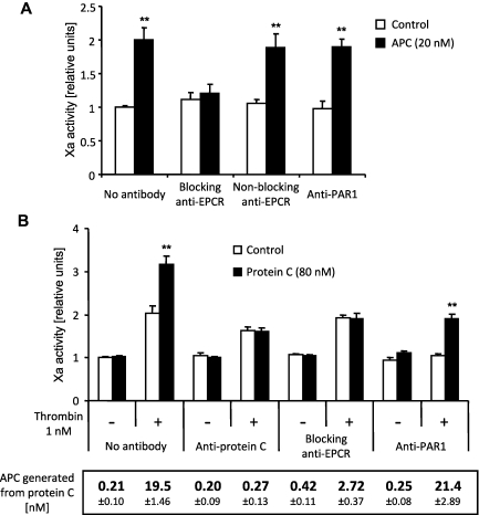
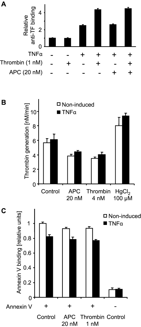
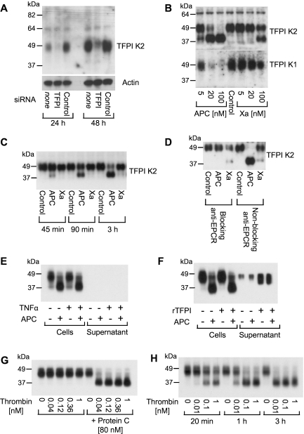
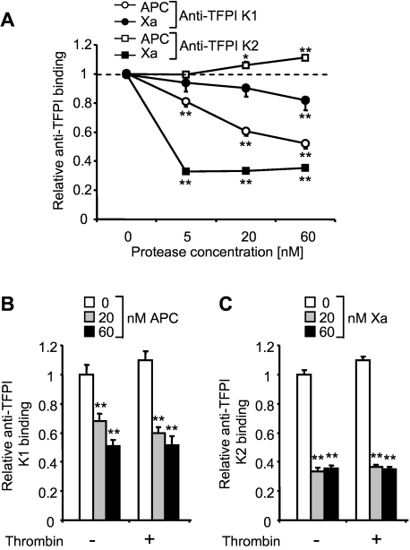
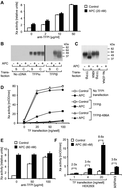
Thrombin and activated protein C (APC) signaling can mediate opposite biologic responses in endothelial cells. Given that thrombin induces procoagulant tissue factor (TF), we examined how TF activity is affected by APC. Exogenous or endogenously generated APC led to increased TF-dependent factor Xa activity. Induction required APC's proteolytic activity and binding to endothelial cell protein C receptor but not protease activated receptors. APC did not affect total TF antigen expression or the availability of anionic phospholipids on the apical cell membrane. Western blotting and cell surface immunoassays demonstrated that APC sheds the Kunitz 1 domain from tissue factor pathway inhibitor (TFPI). A TFPI Lys86Ala mutation between the Kunitz 1 and 2 domains eliminated both cleavage and the enhanced TF activity in response to APC in overexpression studies, indicating that APC up-regulates TF activity by endothelial cell protein C receptor-dependent shedding of the Kunitz 1 domain from membrane-associated TFPI. Our results demonstrate an unexpected procoagulant role of the protein C pathway that may have important implications for the regulation of TF- and TFPI-dependent biologic responses and for fine tuning of the hemostatic balance in the vascular system.
Figures
Similar articles
-
The effect of leukocyte elastase on tissue factor pathway inhibitor.Blood. 1992 Apr 1;79(7):1712-9. Blood. 1992. PMID: 1558967
-
Human tissue factor pathway inhibitor fused to CD4 binds both FXa and TF/FVIIa at the cell surface.Thromb Haemost. 1997 Dec;78(6):1488-94. Thromb Haemost. 1997. PMID: 9423800
-
Ligand-induced protease receptor translocation into caveolae: a mechanism for regulating cell surface proteolysis of the tissue factor-dependent coagulation pathway.J Cell Biol. 1996 Apr;133(2):293-304. doi: 10.1083/jcb.133.2.293. J Cell Biol. 1996. PMID: 8609163 Free PMC article.
-
Structure and biology of tissue factor pathway inhibitor.Thromb Haemost. 2001 Oct;86(4):959-72. Thromb Haemost. 2001. PMID: 11686353 Review.
-
Regulation of coagulation by tissue factor pathway inhibitor: Implications for hemophilia therapy.J Thromb Haemost. 2022 Jun;20(6):1290-1300. doi: 10.1111/jth.15697. Epub 2022 Mar 27. J Thromb Haemost. 2022. PMID: 35279938 Free PMC article. Review.
Cited by
-
TFPI1 mediates resistance to doxorubicin in breast cancer cells by inducing a hypoxic-like response.PLoS One. 2014 Jan 28;9(1):e84611. doi: 10.1371/journal.pone.0084611. eCollection 2014. PLoS One. 2014. PMID: 24489651 Free PMC article.
-
Characterization of Skin Aging-Associated Secreted Proteins (SAASP) Produced by Dermal Fibroblasts Isolated from Intrinsically Aged Human Skin.J Invest Dermatol. 2015 Aug;135(8):1954-1968. doi: 10.1038/jid.2015.120. Epub 2015 Mar 27. J Invest Dermatol. 2015. PMID: 25815425
-
Endothelial Protein C Receptor (EPCR), Protease Activated Receptor-1 (PAR-1) and Their Interplay in Cancer Growth and Metastatic Dissemination.Cancers (Basel). 2019 Jan 8;11(1):51. doi: 10.3390/cancers11010051. Cancers (Basel). 2019. PMID: 30626007 Free PMC article. Review.
-
PAR1 agonists stimulate APC-like endothelial cytoprotection and confer resistance to thromboinflammatory injury.Proc Natl Acad Sci U S A. 2018 Jan 30;115(5):E982-E991. doi: 10.1073/pnas.1718600115. Epub 2018 Jan 17. Proc Natl Acad Sci U S A. 2018. PMID: 29343648 Free PMC article.
-
Factor VII-activating protease promotes the proteolysis and inhibition of tissue factor pathway inhibitor.Arterioscler Thromb Vasc Biol. 2012 Feb;32(2):427-33. doi: 10.1161/ATVBAHA.111.238394. Epub 2011 Nov 23. Arterioscler Thromb Vasc Biol. 2012. PMID: 22116096 Free PMC article.
References
-
- Nemerson Y. The tissue factor pathway of blood coagulation. Semin Hematol. 1992;29(3):170–176. - PubMed
-
- Bach RR. Tissue factor encryption. Arterioscler Thromb Vasc Biol. 2006;26(3):456–461. - PubMed
-
- Broze GJ., Jr Tissue factor pathway inhibitor. Thromb Haemost. 1995;74(1):90–93. - PubMed
-
- Crawley JT, Lane DA. The haemostatic role of tissue factor pathway inhibitor. Arterioscler Thromb Vasc Biol. 2008;28(2):233–242. - PubMed
-
- Esmon CT. The protein C pathway. Chest. 2003;124(3 suppl):26S–32S. - PubMed
Publication types
MeSH terms
Substances
Grants and funding
LinkOut - more resources
Full Text Sources
Miscellaneous
