. 2011 Apr 26;108(17):6975-80.
doi: 10.1073/pnas.1014579108. Epub 2011 Apr 7.

Membrane texture induced by specific protein binding and receptor clustering: active roles for lipids in cellular function

Affiliations

Membrane texture induced by specific protein binding and receptor clustering: active roles for lipids in cellular function

E B Watkins et al. Proc Natl Acad Sci U S A. .

Abstract

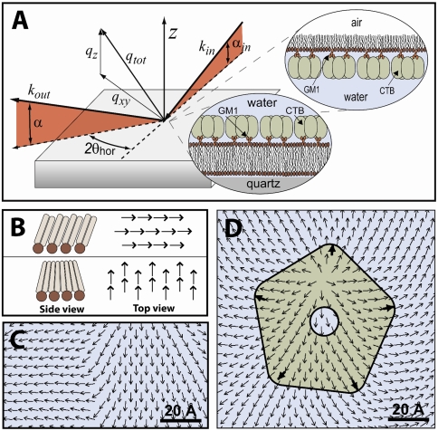
Biological membranes are complex, self-organized structures that define boundaries and compartmentalize space in living matter. Composed of a wide variety of lipid and protein molecules, these responsive surfaces mediate transmembrane signaling and material transport within the cell and with its environment. It is well known that lipid membrane properties change as a function of composition and phase state, and that protein-lipid interactions can induce changes in the membrane's properties and biochemical response. Here, molecular level changes in lipid organization induced by multivalent toxin binding were investigated using grazing incidence X-ray diffraction. Structural changes to lipid monolayers at the air-water interface and bilayers at the solid-water interface were studied before and after specific binding of cholera toxin to membrane embedded receptors. At biologically relevant surface pressures, protein binding perturbed lipid packing within monolayers and bilayers resulting in topological defects and the emergence of a new orientationally textured lipid phase. In bilayers this altered lipid order was transmitted from the receptor laden exterior membrane leaflet to the inner leaflet, representing a potential mechanism for lipid mediated outside-in signaling by multivalent protein binding. It is further hypothesized that cell-surface micro-domains exhibiting this type of lipid order may serve as nucleation sites for vesicle formation in clathrin independent endocytosis of cholera toxin.


Conflict of interest statement

The authors declare no conflict of interest.

Figures

Fig. 1.
Fig. 1.
(A) The GIXD scattering geometry is shown with schematic insets representing the monolayer and bilayer lipid-CTB systems studied. qz = 2π sin α is the momentum transfer of diffracted X-rays normal to the interface and qxy = 4π sin θ is the momentum transfer perpendicular to the interface. (B) Tilt directors are vectors pointing along the lipids’ alkyl chain backbones from the head group to the methyl end. (C) A boundary between two orientations of the lipid tilt director field. (D) Perturbation to the lipid tilt director field and associated topological defect induced by pentavalent binding of a single CTB protein.
Fig. 2.
Fig. 2.
Grazing incidence diffraction from 80∶20 DPPE∶GM1 monolayers at three surface pressures (A1, B1, and C1) and diffraction from the same systems following binding of CTB (A2, B2, and C2). At high surface pressures, protein binding causes an increase in the lipid APM that is commensurate with the increase in lipid tilt. Although APM increased after CTB binding at 20 mN/m, the lipid tilt remained approximately the same. The resulting lipid order was no longer close packed and exhibited topological defects and texture of the lipid tilt orientations.
Fig. 3.
Fig. 3.
Grazing incidence diffraction from monolayers with bound CTB (top) and bilayers before and after CTAB binding (bottom). Diffraction data from the monolayer-CTB complex (A) was reproduced by a textured lipid phase obtained via Monte Carlo simulation (B). (C) Bragg rod profiles extracted from (A) by integrating along qxy and fits corresponding to defect textures of strengths (s = 1, s = 2, s = 3, and s = 5) and the Monte Carlo generated domain (dark line). For a domain radius of 150 Å (corresponding to the measured Bragg Peak FWHM), the scattered intensity at low qz decreased with increasing s. The best fit to the data was found for s ≈ 5. In the bilayer case prior to protein binding (D, F), the Bragg rod FWHM indicates coupling between the membrane leaflets and exhibited limited texture. Following protein binding (E, G), cross leaflet coupling was preserved and the decreased intensity at low qz reflects an increased degree of texture.
Fig. 4.
Fig. 4.
Real space configurations of lipid tilt directors (vectors along the molecular backbones) exhibiting texture. Top left and center schematics show orientation of tilt directors around an s = 1, ϕ0 = π/4 and an s = 5, ϕ0 = π/4 disclination respectively. In the bottom left schematic, ∼10–15 CTB proteins are arranged beneath a 150 Å radius nano-domain corresponding to the lateral correlation size determined from the FWHM of the Bragg Peak. Magnification to the right displays the orientation of tilt directors obtained from Monte Carlo simulation. Dark arrows represent molecules with fixed orientations. A topological defect (source) can be seen near the central pore of the top left CTB molecule.

References

    1. White SH, Wimley WC. Membrane protein folding and stability: Physical principles. Annu Rev Bioph Biom. 1999;28:319–365. - PubMed
    1. Phillips R, et al. Emerging roles for lipids in shaping membrane-protein function. Nature. 2009;459:379–385. - PMC - PubMed
    1. Dietrich C, et al. Lipid rafts reconstituted in model membranes. Biophys J. 2001;80:1417–1428. - PMC - PubMed
    1. Risselada HJ, Marrink SJ. The molecular face of lipid rafts in model membranes. Proc Natl Acad Sci USA. 2008;105:17367–17372. - PMC - PubMed
    1. Simons K, Ikonen E. Functional rafts in cell membranes. Nature. 1997;387:569–572. - PubMed

Publication types

MeSH terms

LinkOut - more resources