Review
. 2011 Sep;18(9):1450-6.
doi: 10.1038/cdd.2011.31. Epub 2011 Apr 8.

The role of PML in the control of apoptotic cell fate: a new key player at ER-mitochondria sites

Affiliations
Review

The role of PML in the control of apoptotic cell fate: a new key player at ER-mitochondria sites

P Pinton et al. Cell Death Differ. 2011 Sep.

Abstract

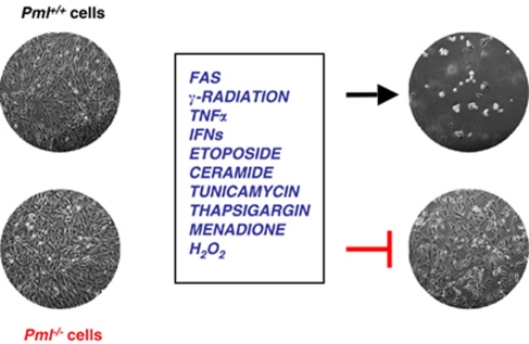
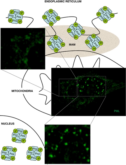
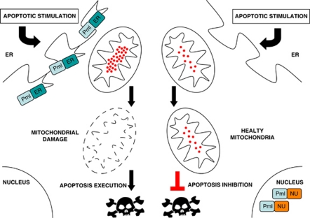
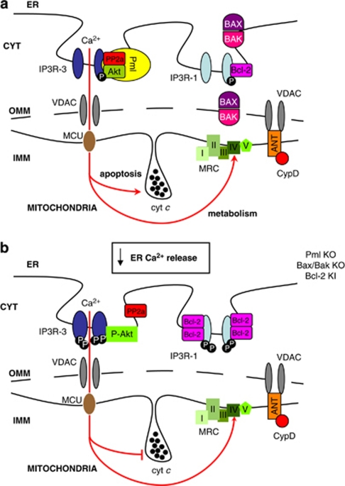
The development of malignant tumors results from deregulated proliferation or an inability of cells to undergo apoptotic cell death. Experimental works of the past decade have highlighted the importance of calcium (Ca(2+)) in the regulation of apoptosis. Several studies indicate that the Ca(2+) content of the endoplasmic reticulum (ER) determines the cell's sensitivity to apoptotic stress and perturbation of ER Ca(2+) homeostasis appears to be a key component in the development of several pathological situations. Sensitivity to apoptosis depends on the ability of cells to transfer Ca(2+) from the ER to the mitochondria. The physical platform for the interplay between the ER and mitochondria is a domain of the ER called the mitochondria-associated membranes (MAMs). The disruption of these contact sites has profound consequences for cellular function, such as imbalances of intracellular Ca(2+) signaling, cellular stress, and disrupted apoptosis progression. The promyelocytic leukemia (PML) protein has been previously recognized as a critical and essential regulator of multiple apoptotic response. Nevertheless, how PML would exert such broad and fundamental role in apoptosis remained for long time a mystery. In this review, we will discuss how recent results demonstrate that the elusive mechanism whereby the PML tumor suppressor exerts its essential role in apoptosis triggered by Ca(2+)-dependent stimuli can be attributed to its unexpected and fundamental role at MAMs in the control of the functional cross-talk between ER and mitochondria.


Figures

Figure 1
Figure 1
PML is critical in multiple apoptotic pathways. The absence of PML inhibits cell death induced by various apoptotic stimuli. PML is critical for both transcription-dependent (e.g., p53-mediated responses) responses as well as transcription-independent early apoptotic responses
Figure 2
Figure 2
Intracellular localization of PML. The PML protein accumulates in the nucleus where it forms PML-nuclear bodies (PML-NBs), as well as at endoplasmic reticulum (ER) in the contact sites with mitochondria (MAMs). In both sites, PML interacts with several proteins (indicated in figure as IP: interacting proteins) generating multi-protein complexes of large molecular size
Figure 3
Figure 3
PML localization at ER/MAM is critical for calcium-dependent apoptotic cell death. Re-expression of an erPML chimera does rescue the sensitivity to cell death of PML null cells. In this case, the ER Ca2+ release induced by various apoptotic stimuli causes a mitochondrial Ca2+ overload and in turn the damage of the mitochondrial structure with release of apoptotic factors. On the contrary, the re-introduction of a nuPML chimera is able to rescue the PML-NBs formation, but not the sensitivity to apoptosis. In this case, the absence of PML at the ER/MAMs sites still impairs ER-Ca2+ release and in turn mitochondrial damage and the apoptotic response
Figure 4
Figure 4
Schematic representation of ER–mitochondria Ca2+ cross-talk perturbations in physiological and oncogenic states. (a) In normal cells, the PML protein is in protein complexes with the type 3 inositol trisphosphate receptor (IP3R-3), the protein kinase Akt and the phosphatase PP2a. This complex is fundamental to regulate the phosphorylation state of IP3R-3, while the Bcl-2-Bax ratio controls the phosphorylation state of the type 1 of IP3R (IP3R-1). In normal conditions, the stimulation of ER Ca2+ release through IP3 generating stimuli allows a transient mitochondrial Ca2+ uptake and in turn the stimulation of mitochondrial ATP production through the mitochondrial respiratory chain (MRC). In contrast, a Ca2+-dependent pro-apoptotic stimulus induces a much larger Ca2+ release from the ER with a dramatic effect on mitochondria, which induces the release of cytocrome c and the opening of the mitochondrial permeability transition pore. This chain of event, combined with the activation of cytoplasmic effectors, is an important trigger of the apoptotic program. (b) In cancer cells, where PML is often missing or when the Bcl-2-Bax ratio is abnormal, IP3R-3 and IPR1 are, respectively, hyper-phosphorylated as demonstrated on Pml KO, Bax/Bak KO, or Bcl-2 KI cells. Under these conditions, pro-apoptotic stimuli induce a smaller release of Ca2+ from ER, with minor repercussions on mitochondrial physiology and intracellular Ca2+-modulated processes

References

    1. Pandolfi PP. Oncogenes and tumor suppressors in the molecular pathogenesis of acute promyelocytic leukemia. Hum Mol Genet. 2001;10:769–775. - PubMed
    1. Salomoni P, Pandolfi PP. The role of PML in tumor suppression. Cell. 2002;108:165–170. - PubMed
    1. Salomoni P, Ferguson BJ, Wyllie AH, Rich T. New insights into the role of PML in tumour suppression. Cell Res. 2008;18:622–640. - PubMed
    1. Bernardi R, Pandolfi PP. Structure, dynamics and functions of promyelocytic leukaemia nuclear bodies. Nat Rev Mol Cell Biol. 2007;8:1006–1016. - PubMed
    1. Jensen K, Shiels C, Freemont PS. PML protein isoforms and the RBCC/TRIM motif. Oncogene. 2001;20:7223–7233. - PubMed

Publication types

MeSH terms