

Food signs in radiology
Int J Health Sci (Qassim).
2007 Jan.
Abstract
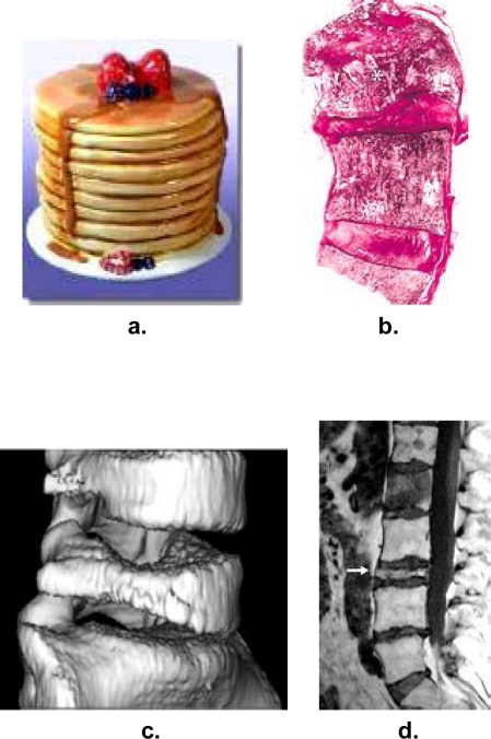
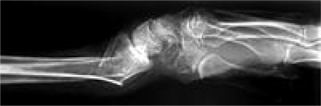
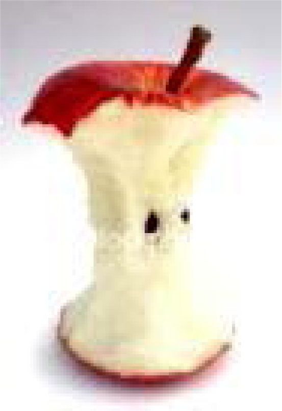
Certain diseases show classic radiological signs that resemble various types of food items like fruits, meat, vegetables, eggs, bakery, grocery and confectionary items. In this article various food signs are discussed and correlated with the various food items in a pictorial way. The objective of this pictorial essay is to provide the information and learn the characteristic radiological signs resembling various food items. These food signs are easy to recognize and allows a confident diagnosis on the basis of imaging findings alone or can narrow down the differential diagnosis.
Keywords: diagnosis; food signs; radiological signs.
Figures


Similar articles
-
Selections from the buffet of food signs in radiology.Radiographics. 2002 Nov-Dec;22(6):1369-84. doi: 10.1148/rg.226025521. Radiographics. 2002. PMID: 12432108 Review.
-
Availability of more healthful food alternatives in traditional, convenience, and nontraditional types of food stores in two rural Texas counties.J Am Diet Assoc. 2009 May;109(5):883-9. doi: 10.1016/j.jada.2009.02.011. J Am Diet Assoc. 2009. PMID: 19394475
-
Pictorial Essay: Classic Signs in Pediatric Neuroradiology.Curr Pediatr Rev. 2020;16(1):6-16. doi: 10.2174/1573396315666190916141023. Curr Pediatr Rev. 2020. PMID: 31526350 Review.
-
Using Variational Multi-view Learning for Classification of Grocery Items.Patterns (N Y). 2020 Nov 13;1(8):100143. doi: 10.1016/j.patter.2020.100143. eCollection 2020 Nov 13. Patterns (N Y). 2020. PMID: 33294874 Free PMC article.
-
[Study of the categorization process among patients with eating disorders: a new cognitive approach to psychopathology].Encephale. 2005 Jan-Feb;31(1 Pt 1):82-91. doi: 10.1016/s0013-7006(05)82376-0. Encephale. 2005. PMID: 15971644 French.
References
-
- Sutton David. 7th ed. Churchill Livingstone; 2002. Text Book of Radiology and Imaging.
-
- Reeder MM. In: Gastrointestinal tract and abdomen Reeder MM. Reeder and Felson’s gamut’s in radiology. 3rd Ed. Bradley WG, editor. New York, NY: Springer-Verlag; 1993.
-
- Dahnert W. Radiology review manual. 3rd ed. Baltimore, MD: Williams & Wilkins; 1996.
-
- Chapman S, Nakielny R. Aids to radiological differential diagnosis. 3rd ed. London, England: Saunders; 1995.
-
- Felson B. A new look at pattern recognition of diffuse pulmonary disease. AJR Am J Roentgenol. 1979;133:183–189. - PubMed
LinkOut - more resources
Full Text Sources