. 2011 Mar 8;4(1):16-22.
doi: 10.1007/s12178-011-9072-0.

Assessment of scaphoid fracture healing

Affiliations

Assessment of scaphoid fracture healing

Lauren A Hackney et al. Curr Rev Musculoskelet Med. .

Abstract

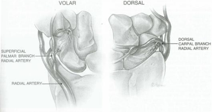
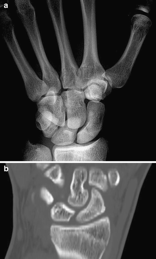
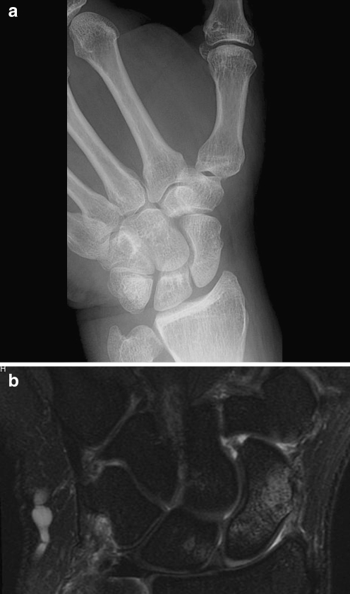
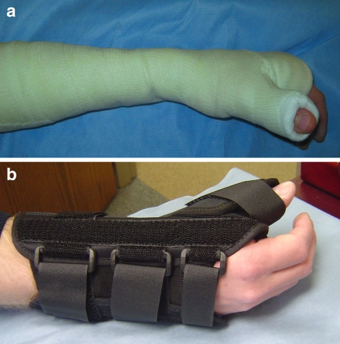
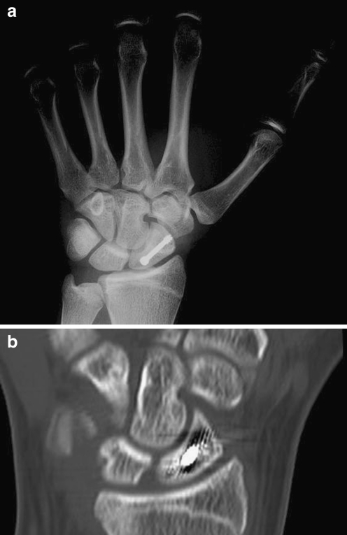
Scaphoid fractures are among the most common hand fractures in adults. The geometry of the scaphoid as it relates to its retrograde blood supply renders it particularly prone to avascular necrosis and other fracture complications. Though there has been long-standing debate over the optimal method of diagnosing scaphoid fractures, the best and most cost-effective methods combine clinical exam with other imaging modalities such as navicular view plain films, CT, and MRI for particularly questionable presentations. Once a scaphoid fracture is diagnosed, it should be followed by an orthopaedic surgeon and treated with cast immobilization or operative management in the case of displaced fractures. Fractures should be followed to monitor healing progress in order to ensure the eventual development of bridging bone across the fracture line, usually best appreciated on CT. Proper treatment of scaphoid fractures and assessment of fracture healing can minimize the occurrence of non-unions and associated arthritic changes.

Keywords: Healing; Imaging; Non-union; Scaphoid fracture.


Figures

Fig. 1
Fig. 1
Blood supply of the scaphoid. With permission from [27].
Fig. 2
Fig. 2
a The snuffbox of the wrist is examined for tenderness by deeply palpating the proximal pole of the scaphoid between the abductor pollicis longus and extensor pollicis brevis tendons radially and the extensor pollicis longus tendon ulnarly, just distal to the radial styloid. b The distal tubercle of the scaphoid is the volar and distal most aspect of this carpal bone and can be palpated at the radial aspect of the distal volar wrist crease at the base of the thumb
Fig. 3
Fig. 3
a Plain posteroanterior radiograph with the wrist in ulnar deviation to bring the scaphoid into a profile of extension barely demonstrates a fracture line. b CT scan image of the same patient reformatted along the axis of the scaphoid more clearly reveals the fracture line
Fig. 4
Fig. 4
a X-ray of the wrist is completely normal on navicular view. b MRI of the same patient shows increased inflammation and edema at the scaphoid waist, indicative of a fracture
Fig. 5
Fig. 5
a Short arm thumb spica cast extends from the proximal forearm to the thumb, immobilizing the wrist and the thumb ray (which extends off the distal pole of the scaphoid) to provide external stabilization of the scaphoid. b Thumb spica brace
Fig. 6
Fig. 6
a Plain film of a patient status post surgical treatment with scaphoid screw. b CT of same patient showing confirmed healing

References

    1. Gaebler C, McQueen MM. Carpus fractures and dislocations. In: Bucholz RW, Heckman JD, Court-Brown CM, Tornetta P, editors. Fractures in adults. 3. Philadelphia: Lippincott Williams and Wilkins; 2009. pp. 782–828.
    1. Barton N. Twenty questions about scaphoid fractures. J Hand Surg Br. 1992;17:289–310. doi: 10.1016/0266-7681(92)90118-L. - DOI - PubMed
    1. Ring D, Jupiter J, Herndon J. Acute fractures of the scaphoid. J Am Acad Orthop Surg. 2000;8:225–231. - PubMed
    1. Waizenegger M, Barton N, Davis T, Wastie M. Clinical signs in scaphoid fractures. J Hand Surg Br. 1994;19:743–747. doi: 10.1016/0266-7681(94)90249-6. - DOI - PubMed
    1. Parvizi J, Wayman J, Kelly P, Moran C. Combining the clinical signs improves diagnosis of scaphoid fractures. A prospective study with follow-up. J Hand Surg Br. 1998;23:324–327. doi: 10.1016/S0266-7681(98)80050-8. - DOI - PubMed