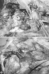
Successful function-preserving therapy for chondroblastoma of the temporal bone involving the temporomandibular joint
- PMID: 21475594
- PMCID: PMC3072183
- DOI: 10.1159/000324640
Successful function-preserving therapy for chondroblastoma of the temporal bone involving the temporomandibular joint
Abstract
We present a case involving a late diagnosis of chondroblastoma of the temporal skull base involving the temporomandibular joint (TMJ). Following an initial misdiagnosis and unsuccessful treatment over a period of 5 years, the patient was referred to our department for further evaluation and possible surgical intervention for occlusal abnormalities, trismus, clicking of the TMJ, and hearing impairment. Based on preoperative immunochemical studies showing positive reaction of multinucleated giant cells for S-100 protein, the final diagnosis was chondroblastoma. The surgical approach - postauricular incision and total parotidectomy, with complete removal of the temporal bone, including the TMJ via the extended middle fossa - was successful in preserving facial nerves and diminishing clinical manifestations. This study highlights a misdiagnosed case in an effort to underline the importance of medical examinations and accurate differential diagnosis in cases involving any tumor mass in the temporal bone.
Keywords: Chondroblastoma; Parotidectomy; Temporal bone; Temporomandibular joint; Trismus.
Figures


Similar articles
-
Chondroblastoma of the temporal bone involving the temporomandibular joint, mandibular condyle, and middle cranial fossa: case report and review of the literature.Cranio. 2004 Apr;22(2):160-8. doi: 10.1179/crn.2004.021. Cranio. 2004. PMID: 15134417 Review.
-
Tenosynovial giant cell tumors of the temporomandibular joint and lateral skull base: Review of 11 cases.Laryngoscope. 2017 Oct;127(10):2340-2346. doi: 10.1002/lary.26435. Epub 2016 Nov 26. Laryngoscope. 2017. PMID: 27888510
-
Surgical treatment and outcomes of temporal bone chondroblastoma.Eur Arch Otorhinolaryngol. 2008 Dec;265(12):1447-54. doi: 10.1007/s00405-008-0660-6. Epub 2008 Apr 10. Eur Arch Otorhinolaryngol. 2008. PMID: 18401591
-
Chondroblastoma of the temporal bone.Acta Otolaryngol. 2011 Aug;131(8):890-5. doi: 10.3109/00016489.2011.566579. Epub 2011 Apr 19. Acta Otolaryngol. 2011. PMID: 21504272
-
Temporal Bone Chondroblastoma: Systematic Review of Clinical Features and Outcomes.World Neurosurg. 2020 Oct;142:e260-e270. doi: 10.1016/j.wneu.2020.06.192. Epub 2020 Jun 27. World Neurosurg. 2020. PMID: 32603862
Cited by
-
Functional treatment of temporal bone chondroblastoma: retrospective analysis of 3 cases.Eur Arch Otorhinolaryngol. 2021 Apr;278(4):1271-1276. doi: 10.1007/s00405-020-06203-4. Epub 2020 Jul 13. Eur Arch Otorhinolaryngol. 2021. PMID: 32661717
-
Chondroblastoma of mandibular condyle: Case report and literature review.Open Med (Wars). 2021 Sep 13;16(1):1372-1377. doi: 10.1515/med-2021-0352. eCollection 2021. Open Med (Wars). 2021. PMID: 34595350 Free PMC article.
-
Variants and Modifications of the Retroauricular Approach Using in Temporomandibular Joint Surgery: A Systematic Review.J Clin Med. 2021 May 11;10(10):2049. doi: 10.3390/jcm10102049. J Clin Med. 2021. PMID: 34064639 Free PMC article. Review.
-
A large invasive chondroblastoma on the temporomandibular joint and external auditory canal: a case report and literature review.Maxillofac Plast Reconstr Surg. 2021 Jul 14;43(1):26. doi: 10.1186/s40902-021-00313-7. Maxillofac Plast Reconstr Surg. 2021. PMID: 34259979 Free PMC article.
References
-
- Dahlin DS, Ivins JC. Benign chondroblastoma. A study of 125 cases. Cancer. 1972;30:401–413. - PubMed
-
- Denko JV, Krauel LH. Benign chondroblastoma of bone: an unusual localization in temporal bone. AMA Arch Path. 1955;59:710–711. - PubMed
-
- Hamer SG, Cody DT, Dahlin DC. Benign chondrosarcoma of the temporal bone. Otolaryngol Head Neck Surg. 1979;87:229–236. - PubMed
-
- Steiner GC. Postradiation sarcoma of bone. Cancer. 1965;18:603–612. - PubMed
Publication types
LinkOut - more resources
Full Text Sources

