. 2008 Dec;36(7):142s-163s.
doi: 10.1177/0192623308327117.

Selected Background Findings and Interpretation of Common Lesions in the Female Reproductive System in Macaques

Affiliations

Selected Background Findings and Interpretation of Common Lesions in the Female Reproductive System in Macaques

J Mark Cline et al. Toxicol Pathol. 2008 Dec.

Abstract

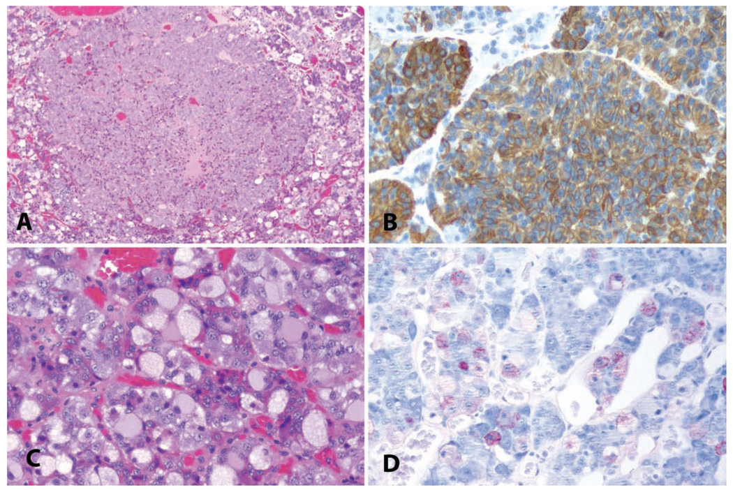
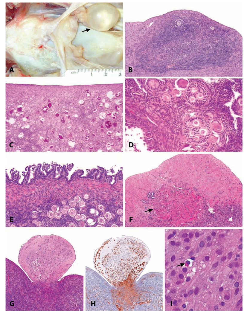
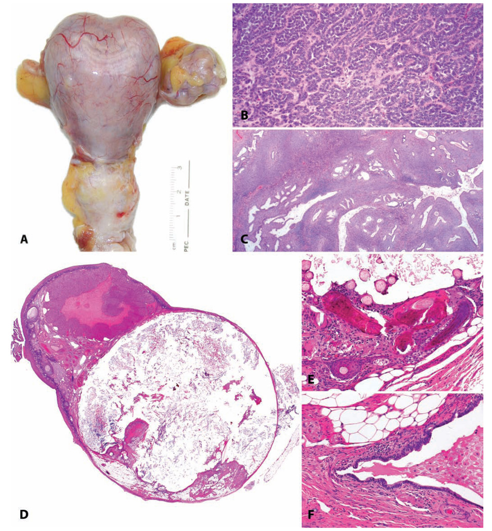
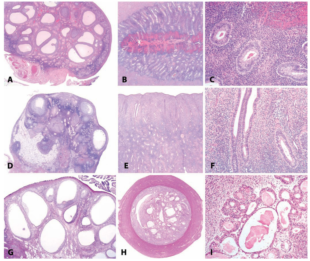
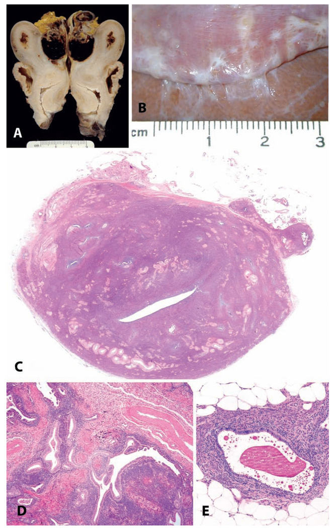
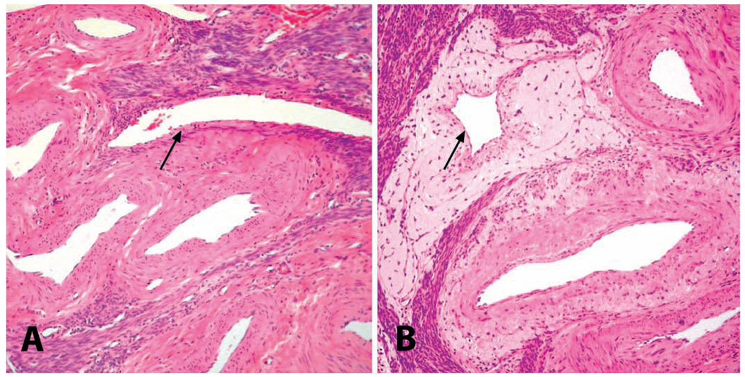
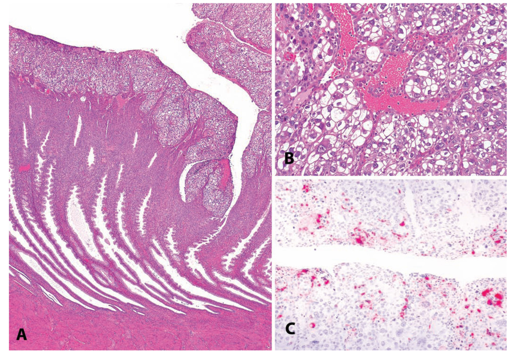
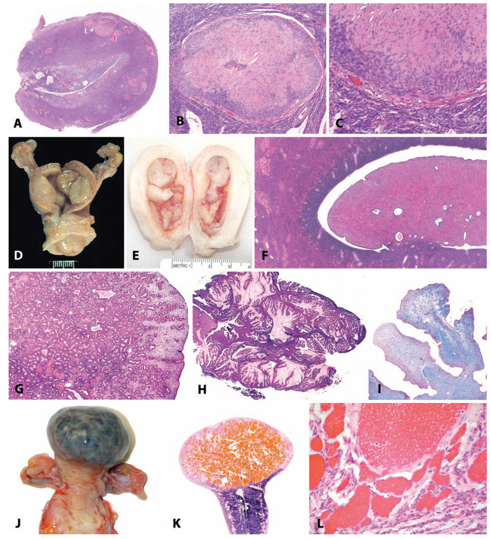
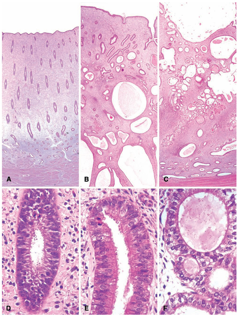
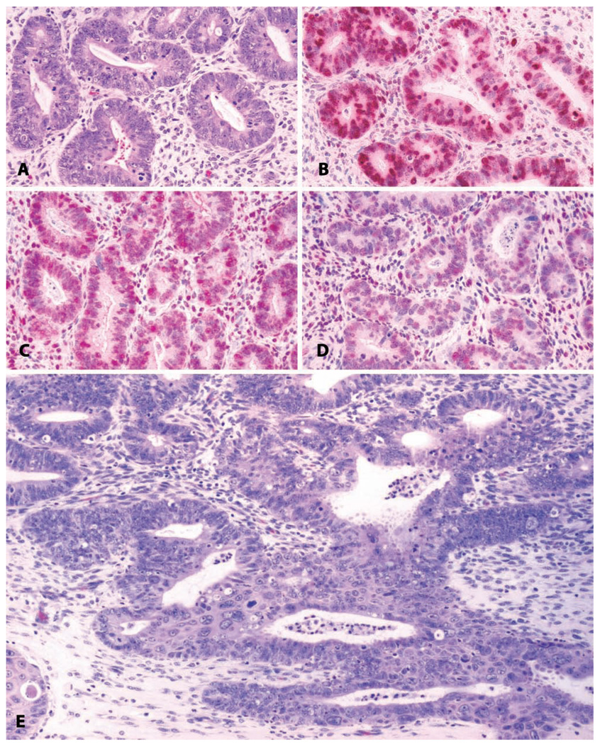
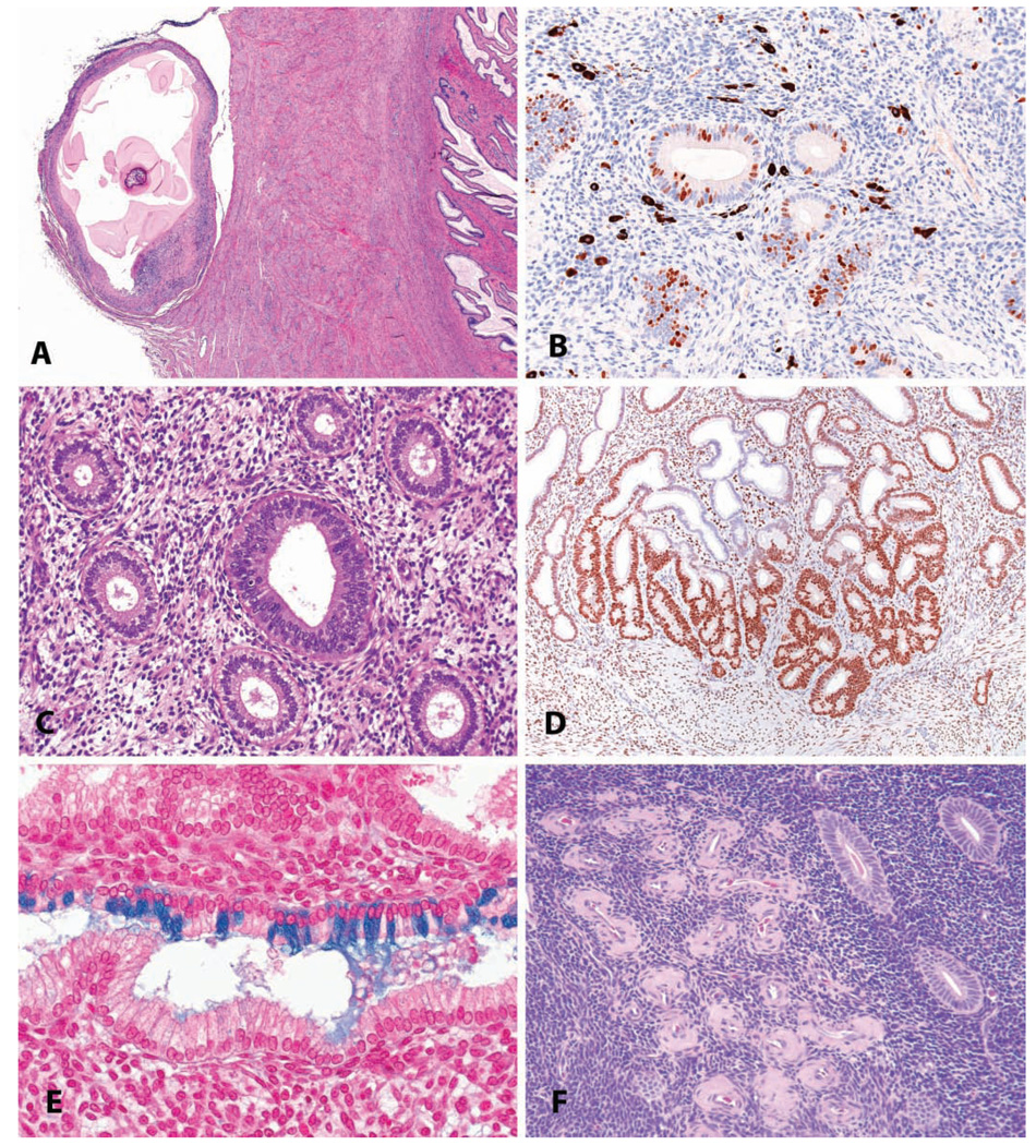
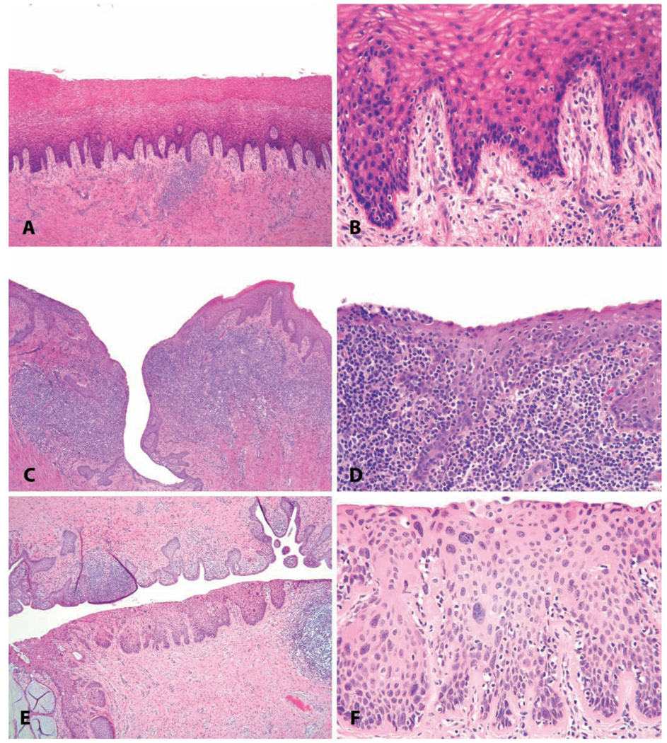
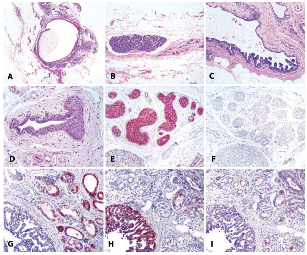
The authors describe a selection of normal findings and common naturally occurring lesions in the reproductive system of female macaques, including changes in the ovaries, uterus, cervix, vagina, and mammary glands. Normal features of immature ovaries, uteri, and mammary glands are described. Common non-neoplastic lesions in the ovaries include cortical mineralization, polyovular follicles, cysts, ovarian surface epithelial hyperplasia, and ectopic ovarian tissue. Ovarian neoplasms include granulosa cell tumors, teratomas, and ovarian surface epithelial tumors. Common non-neoplastic uterine findings include loss of features of normal cyclicity, abnormal bleeding, adenomyosis, endometriosis, epithelial plaques, and pregnancy-associated vascular remodeling. Hyperplastic and neoplastic lesions of the uterus include endometrial polyps, leiomyomas, and rarely endometrial hyperplasia and endometrial adenocarcinoma. Vaginitis is common. Cervical lesions include endocervical squamous metaplasia, polyps, and papillomavirus-associated lesions. Lesions in the mammary gland are most often proliferative and range from ductal hyperplasia to invasive carcinoma. Challenges to interpretation include the normal or pathologic absence of menstrual cyclicity and the potential misinterpretation of sporadic lesions, such as epithelial plaques or papillomavirus-associated lesions. Interpretation of normal and pathologic findings is best accomplished with knowledge of the life stage, reproductive history, and hormonal status of the animal.


Figures

FIGURE 1
FIGURE 1
(A) Pituitary microadenoma in an aged cynomolgus macaque. H&E. (B) Pituitary adenoma in a twenty-five-year-old cynomolgus macaque. Immunohistochemistry for adrenocorticotropic hormone; hematoxylin counterstain. (C) Gonadotroph hypertrophy in the pars distalis of an adult ovariectomized cynomolgus macaque. H&E. (D) Follicle-stimulating hormone immunostain, hematoxylin counterstain.
FIGURE 2
FIGURE 2
(A) Gross appearance of a para-ovarian cyst in a nineteen-year-old cynomolgus monkey. (B) Ectopic ovarian tissue on the uterine serosal surface of an adult, normally cycling cynomolgus monkey. Note the presence of primary and atretic follicles in a dense ovarian-type stroma. H&E. (C) Multifocal mineralization in the ovary of a 2.5-year-old cynomolgus monkey. H&E. (D) Polyovular follicles in the ovary of a 2.5-year-old cynomolgus monkey. H&E. (E) Papillary hyperplasia of the ovarian surface epithelium in a 7.5-year-old cynomolgus monkey. H&E. (F) Focal smooth muscle metaplasia of the ovarian stroma in the ovary of an 18-year-old cynomolgus monkey; the absence of oocytes and fibrosis of the adjacent stroma reflects the age of the animal. H&E. (G, H, I) Ovarian deciduosis in a 16-year-old cynomolgus monkey, forming a polypoid mass projecting above the ovarian surface. (G) H&E. (H) anti-smooth muscle actin. (I) H&E at higher magnification. Typical endometrial lymphocytes were present (arrow).
FIGURE 3
FIGURE 3
Ovarian neoplasia. (A–C) Granulosa cell tumor from a twenty-six-year-old cynomolgus monkey with an estrogen-producing ovarian granulosa cell tumor, with the functional consequence of uterine enlargement by endometrial hyperplasia and myometrial hypertrophy. (A) Gross specimen. The uterus weighed more than 70 grams, compared to <2 grams for an ovariectomized animal or 7 grams for a cycling animal. (B) Typical histologic appearance of ovarian granulosa cell tumors. H&E. (C) Simple endometrial hyperplasia induced by estrogens of tumoral origin. H&E. (D–F) Benign ovarian teratoma in an 8.5-year-old cynomolgus monkey. (D) Subgross histologic appearance of this cystic neoplasm. H&E. (E, F) Multiple tissue types including hair, bone, fat, and glandular epithelium. H&E.
FIGURE 4
FIGURE 4
Three syndromes of ovary-dependent dysregulation of the endometrium. (A–C) Abnormal endometrial bleeding in a pubertal macaque; note the absence of a corpus luteum (A) and hemorrhage in the functionalis without prior luteal differentiation (B). In panel (C), the absence of subnuclear vacuoles or stromal decidualization indicate that the bleeding is out of cycle. (D–F) Suppression of ovarian function in a 21.5-year-old cynomolgus monkey, with quiescent ovarian and endometrial morphology. (G–I) Polycystic ovarian syndrome in a thirteen-year-old cynomolgus monkey, characterized by polycystic ovaries and endometrial hyperplasia. This pattern should not be confused with (A).
FIGURE 5
FIGURE 5
Endometriosis and adenomyosis. (A and B) Variation in the gross appearance of endometriosis in macaques. (A) Typical blood-filled cyst caudal to the uterus in an adult female cynomolgus monkey. (B) Plaque-like pale lesions causing adhesions between the liver and diaphragm in a nineteen-year-old cynomolgus monkey. (C) Transverse section of the uterus of a twenty-four-year-old cynomolgus monkey with both adenomyosis and endometriosis, causing asymmetry of the uterus. H&E. (D and E) Typical histologic appearance of endometriosis, including endometrial glands, stroma, and hemorrhage or hemosiderin. H&E.
FIGURE 6
FIGURE 6
Uteri of adult cynomolgus monkeys. Myometrial vasculature of (A) a nulliparous animal and (B) a multiparous animal, demonstrating the perivenous accumulation of loose extracellular matrix indicative of prior pregnancy. (A) Myometrial vasculature of a nulliparous animal; note the nearly invisible wall of myometrial vein (arrow). (B) Similar area from a multiparous animal, showing perivascular accumulation of pale eosinophilic extracellular matrix surrounding a myometrial vein (arrow). H&E.
FIGURE 7
FIGURE 7
An epithelial plaque in a 3.5-year-old cynomolgus monkey. (A) Low-magnification photomicrograph demonstrating the surface orientation and concurrent endometrial glandular features of the luteal phase. (B) Higher magnification. H&E. (C) Immunohistochemistry for cleaved caspase 3, indicating apoptosis, in a regressing plaque. Vector red chromogen; hematoxylin counterstain.
FIGURE 8
FIGURE 8
Benign uterine neoplasms. (A) Longitudinal section through the uterus of a thirty-year-old rhesus macaque; within the myometrium there are four leiomyomas. H&E. (B and C) Higher magnification demonstrating smooth muscle morphology of the neoplastic cells. H&E. (D and E) Gross photographs of endometrial polyps in aged rhesus macaques; the polyps in (D) are sessile, and those in (E) are pedunculated. (F) Endometrial polyp consisting primarily of stromal tissue in a fourteen-year-old cynomolgus monkey. H&E. (G) Endometrial polyp in an adult rhesus monkey consisting primarily of glandular tissue. H&E. (H) Endometrial polyp in an adult rhesus monkey, with mucous differentiation. H&E. (I) Fibrosis within a polyp in an adult cynomolgus monkey; Masson’s trichrome stain. (J) Gross photograph of a myometrial cavernous hemangioma in a twenty-two-year-old cynomolgus monkey. (K) Subgross appearance. (L) Higher magnification. H&E.
FIGURE 9
FIGURE 9
Uterus: normal and pathologic proliferative changes. (A and D) Normal follicular phase endometrium with edema of the functionalis and orderly glandular proliferation. (B and E) Simple endometrial hyperplasia with disorganized glandular structures, cystic change, and ciliary metaplasia of the glandular epithelium. (C and F) Complex endometrial hyperplasia, with “back-to-back” crowding of endometrial glands.
FIGURE 10
FIGURE 10
Uterus: Hyperplasia with progression to invasive endometrial carcinoma. (A) Endometrial glandular hyperplasia in a thirty-two-year-old rhesus macaque chronically treated with estradiol by subcutaneous implant. “Complex hyperplasia” or “endometrial intraepithelial neoplasia.” H&E. (B) Corresponding area stained for proliferating cells; anti-Ki-67 antibody, Vector red chromogen, and hematoxylin counterstain. (C) Corresponding area stained for estrogen receptor alpha; anti-ER antibody, Vector red chromogen, and hematoxylin counterstain. (D) Corresponding area stained for progesterone receptors; anti-PR antibody, Vector red chromogen, and hematoxylin counterstain. (E) Adjacent region of the same slide; highly pleomorphic and invasive endometrial adenocarcinoma. H&E.
FIGURE 11
FIGURE 11
Uterus: incidental findings. (A) Parasitic remnant manifest as a fibrous/mineralized cyst on the cervical serosa in a four-year-old cynomolgus monkey. H&E. (B) Endometrial melanosis in a four-year-old cynomolgus monkey, shown in an immunohistochemical stain for the proliferation marker. Immunohistochemistry: diaminobenzidine chromogen, hematoxylin counterstain. (C) “Luteal phase defect” in an adult cynomolgus monkey, with a mixture of follicular-phase features (round glandular lumens, pseudostratification) and luteal phase features (stro-mal proliferation and hypertrophy, glandular subnuclear vacuoles). (D) Focal glandular hyperplasia, basalis, in an adult cynomolgus monkey. Progesterone receptor immunostain, diaminobenzidine chromogen, hematoxylin counterstain. (E). Endometrial glandular mucous metaplasia in an adult cynomolgus monkey. Alcian blue stain. (F) Focal endometrial hyaline perivascular deposits in an aged cynomolgus monkey. H&E.
FIGURE 12
FIGURE 12
Common vaginal lesions. (A–D) Vaginitis. (A and B) Minimal lymphocytic vaginitis, in a 22.5-year-old intact cycling animal with vaginal keratinization. (C and D) Severe chronic vaginitis with lymphoid follicular hyperplasia in a 7.5-year-old ovariectomized cynomolgus monkey (note atrophy of the surface epithelium and lack of keratinization). (E and F) Papillomavirus-induced in situ neoplasic lesion in a twenty-three-year-old cynomolgus monkey (cervical intraepithelial neoplasia, grade 2). H&E.
FIGURE 13
FIGURE 13
Benign and malignant breast lesions. (A) Cystic change in a sixteen-year-old cynomolgus monkey, with marked dilatation of a large duct. H&E. (B) Focal lobular hyperplasia in the mammary gland of an adult cynomolgus monkey. Note the atrophy of other glandular elements on the right side of the photo. H&E. (C) Papillary ductal hyperplasia in an adult cynomolgus monkey. H&E. (D) Ductal hyperplasia with atypia in an eighteen-year-old cynomolgus monkey. H&E. (E and F) The same lesion as in (D) stained for estrogen receptor and the proliferation marker Ki67, respectively. Note overexpression of estrogen receptor. (G, H, and I) Ductal carcinoma in a five-year-old rhesus macaque, immunostained for (G) estrogen receptor, (H) proliferating cells/Ki67, and (I) progesterone receptor. Vector red chromogen; hematoxylin counterstain.

References

    1. Abdel-Fatah TM, Powe DG, Hodi Z, Lee AH, Reis-Filho JS, Ellis IO. High frequency of coexistence of columnar cell lesions, lobular neoplasia, and low grade ductal carcinoma in situ with invasive tubular carcinoma and invasive lobular carcinoma. Am J Surg Pathol. 2007;31:417–426. - PubMed
    1. Adams MR, Kaplan JR, Koritnik DR. Psychosocial influences on ovarian endocrine and ovulatory function in Macaca fascicularis. Physiol Behav. 1985;35:935–940. - PubMed
    1. Ami Y, Suzaki Y, Goto N. Endometriosis in cynomolgus monkeys retired from breeding. J Vet Med Sci. 1993;55:7–11. - PubMed
    1. Arifin E, Shively CA, Register TC, Cline JM. Polycystic ovary syndrome with endometrial hyperplasia in a cynomolgus monkey (Macaca fascicularis) Veterinary Pathology. in press. - PubMed
    1. Ashbeck EL, Rosenberg RD, Stauber PM, Key CR. Benign breast biopsy diagnosis and subsequent risk of breast cancer. Cancer Epidemiol Biomarkers Prev. 2007;16:467–472. - PubMed