

Evaluation of molecular diagnosis in fungal keratitis. Ten years of experience
- PMID: 21475656
- PMCID: PMC3062769
- DOI: 10.1007/s12348-011-0019-9
Evaluation of molecular diagnosis in fungal keratitis. Ten years of experience
Abstract
Purpose: The aims of this study were to assess the utility of polymerase chain reaction (PCR) in diagnosing fungal keratitis in the last decade in our center and to review the molecular diagnosis of mycotic keratitis.
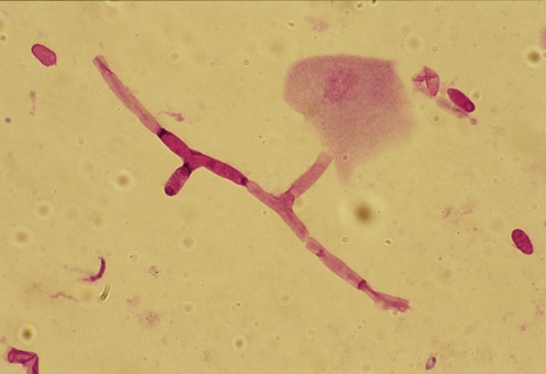
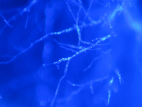
Methods: A retrospective nonrandomized investigation was undertaken at Vissum Corporación Instituto Oftalmologico de Alicante to evaluate 27 corneal samples of 20 patients with proven fungal keratitis from January 2000 to December 2009. Corneal samples (21 corneal scrapings, 5 biopsies, and 1 cornea) were evaluated by Gram stain or calcofluor stain, culture, and PCR. The detection and molecular identification were carried out by DNA amplification and sequencing of the internal transcribed spacer and 5.8S rRNA region from the corneal samples.
Results: PCR detected all the samples that were positive by conventional methods. Four samples were positive by PCR and showed negative results by culture and stain. Combination of microscopy and culture gave positive results in 21 of the 27 samples of patients with mycotic keratitis. Stains showed a 66.7% of positive results, culture showed 59.3%, and PCR showed 92.6%. The time taken for PCR assay was 4 to 8 h whereas positive fungal cultures took 1 to 35 days. Identification at species level by molecular methods was possible in all cases except one. Identification at species level by conventional methods only was possible in eight cases.
Conclusions: PCR not only proved to be an effective rapid method for the diagnosis of fungal keratitis but was also more sensitive than stain and culture methods. Fungal PCR must be added as the screening diagnosis test when an early mycotic keratitis is suspected. Molecular identification is the gold standard technique for the identification of corneal fungal pathogens.
Keywords: Diagnosis; Fungi; Infection; Keratitis; Molecular diagnosis.
Figures
References
-
- Harris DJ, Jr, Stulting RD, Waring GO, III, Wilson LA. Late bacterial and fungal keratitis after corneal transplantation. Spectrum of pathogens, graft survival, and visual prognosis. Ophthalmology. 1988;95:1450–1457. - PubMed
-
- Liesegang TJ, Forster RK. Spectrum of microbial keratitis in South Florida. Am J Ophthalmol. 1980;90:38–47. - PubMed
LinkOut - more resources
Full Text Sources
