Comparative Study
. 2011 May 17;50(19):3879-90.
doi: 10.1021/bi101702c. Epub 2011 Apr 22.

Cardiolipin deficiency in Rhodobacter sphaeroides alters the lipid profile of membranes and of crystallized cytochrome oxidase, but structure and function are maintained

Affiliations
Comparative Study

Cardiolipin deficiency in Rhodobacter sphaeroides alters the lipid profile of membranes and of crystallized cytochrome oxidase, but structure and function are maintained

Xi Zhang et al. Biochemistry. .

Abstract

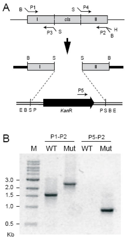
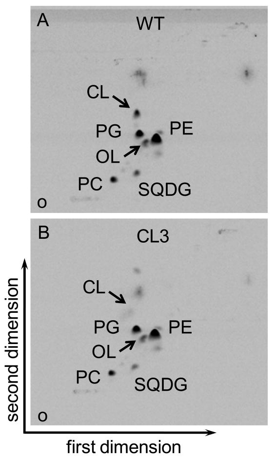
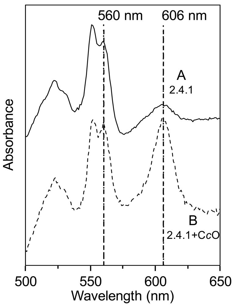
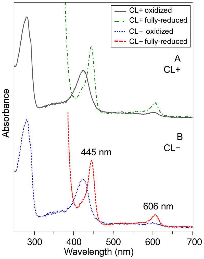
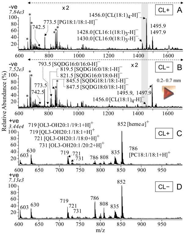
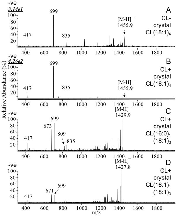
Many recent studies highlight the importance of lipids in membrane proteins, including in the formation of well-ordered crystals. To examine the effect of changes in one lipid, cardiolipin, on the lipid profile and the production, function, and crystallization of an intrinsic membrane protein, cytochrome c oxidase, we mutated the cardiolipin synthase (cls) gene of Rhodobacter sphaeroides, causing a >90% reduction in cardiolipin content in vivo and selective changes in the abundances of other lipids. Under these conditions, a fully native cytochrome c oxidase (CcO) was produced, as indicated by its activity, spectral properties, and crystal characteristics. Analysis by MALDI tandem mass spectrometry (MS/MS) revealed that the cardiolipin level in CcO crystals, as in the membranes, was greatly decreased. Lipid species present in the crystals were directly analyzed for the first time using MS/MS, documenting their identities and fatty acid chain composition. The fatty acid content of cardiolipin in R. sphaeroides CcO (predominantly 18:1) differs from that in mammalian CcO (18:2). In contrast to the cardiolipin dependence of mammalian CcO activity, major depletion of cardiolipin in R. sphaeroides did not impact any aspect of CcO structure or behavior, suggesting a greater tolerance of interchange of cardiolipin with other lipids in this bacterial system.


Figures

Figure 1
Figure 1
Disruption of the cls gene. (A) Schematic diagram showing the gene disruption strategy. Grey boxes represent the cls gene and amplified segments of the gene. The black block arrow represents the kanamycin resistance cassette and the direction of the arrowhead shows its orientation. Plasmids pPICT-2 and pUC4K are shown as thick solid lines and double lines, respectively. Restriction sites: B, BamHI; S, SalI, H, HindIII; E, EcoRI; P, PstI. (B) PCR verification for the gene disruption. Genomic DNA was isolated from the type (WT) and the cls-deleted mutant (Mut) strains and two PCR reactions were carried out using different sets of primers placed at the positions indicated in Fig. 2A. The sizes of the PCR products were compared by gel electrophoresis. M indicates the marker lane.
Figure 2
Figure 2
Two-dimensional thin-layer chromatogram of 14C-labeled lipids from the aerobically-grown R. sphaeroides (A) wild-type strain 2.4.1 and (B) the cls-deleted mutant CL3. The lipids were visualized by using a phosphoimager. Lipids: PE, phosphatidylethanolamine; PG, phosphatidylglycerol; PC, phosphatidylcholine; SQDG, sulfolipid; OL, ornithine lipid; CL, cardiolipin. O, origin.
Figure 3
Figure 3
Growth curves of R. sphaeroides wild-type (▲; 2.4.1) and CL-deficient mutant (●; CL3) strains under A) anaerobic photoheterotrophic growth, and B) aerobic chemoheterotrophic growth conditions, both in Sistrom’s media at 30 °C. The data presented are the average of three independent growth cultures and the error bars are the standard deviations. Absorbances are expressed in Klett units, where 1 Klett unit is equivalent to 107 cells per mL (38).
Figure 4
Figure 4
Optical difference spectra of dithionite-reduced minus ferricyanide-oxidized solubilized membranes from A) R. sphaeroides strains 2.4.1 (wild-type) and B) 2.4.1+CcO (wild-type with CcO genes added on the expression plasmid PRK- pYJ123H). The relative levels of CcO are indicated by the sizes of the a-type heme absorbance peaks at 606 nm compared to the b-heme at 560 nm. The spectra are normalized to the 560 nm (b-type heme) peak. (Note that these membranes also contain the bc1 complex, a cbb3 type oxidase, and a membrane-bound cytochrome c which all contribute to the 560 nm and 550 nm peaks.)
Figure 5
Figure 5
MALDI-MS spectra of membranes isolated from CL(+) (169WT) and CL(−) (169CL3) strains of R. sphaeroides in negative ion mode (A and B) and in positive ion mode (C and D). The highlighted peaks in A) are those of CL at 1456, 1430, and 1428, which are barely discernable in B), the CL(–) sample. A) CL(+) membranes in negative ion mode, B) CL(−) membranes in negative ion mode, C) CL(+) membranes in positive ion mode, D) CL(−) membranes in positive ion mode, each at 0.25 mg/mL membrane protein concentration. The identities of labeled ions in each spectrum were confirmed by MS/MS experiments. Ions labeled with asterisks were identified as 2,5-DHB matrix cluster adducts. The peaks at m/z 1495.9, 1497.9, 1533.8 and 1535.8 were determined by MS/MS and MSn not to be CL or CL derivatives.
Figure 6
Figure 6
Oxidized and reduced spectra of purified CcO samples used to produce protein crystals, purified from A) CL(+) (169WT) and B) CL(−) (169CL3) strains grown aerobically at 30 ºC. The reduced peaks at 445 and 606 nm, which are characteristic of the native heme a and a3 spectra, are seen to be at the same wavelengths in both CcO forms. These peaks have contributions of both hemes at both wavelengths. The spectra were acquired using the same CcO protein concentrations.
Figure 7
Figure 7
MALDI-MS spectra of the four-subunit CcO protein crystals from CL(+) (169WT) and CL(−) (169CL3) strains of R. sphaeroides in negative ion mode (A and B) and positive ion mode (C and D), showing a marked difference in CL content by relative peak height at m/z 1456 (highlighted). A) CcO from CL(+) in negative ion mode, B) CcO from CL(−) in negative ion mode, C) CcO from CL(+) in positive ion mode, D) CcO from CL(−) in positive ion mode. Ions labeled with asterisks were identified as 2,5-DHB matrix cluster adducts. The identities of labeled ions in each spectrum were confirmed by MS/MS experiments. Inset in B: photo of a typical four-subunit CcO crystal used in this analysis.
Figure 8
Figure 8
Identification of the cardiolipin molecular species in the four-subunit CcO crystals from CL(+) (169WT) and CL(−) (169CL3) R. sphaeroides using CID MS/MS in negative ion mode on a MALDI-linear ion trap mass spectrometer, showing the marked depletion of CL(18:1)4 in CL(−) crystals compared to CL(+) crystals (A and B) as demonstrated by comparison of the absolute ion abundance of m/z 699 (top left of panels A and B ), the predominant product ion generated by CL(18:1)4 in MS/MS, corresponding to the phosphatidyldioleoylglyceride moiety. The figure identifies multiple CL species in CL(+) CcO crystals (B, C, D) but no CL (18:2)4 at m/z 1448 is seen (see also Figure 7A).
Figure 9
Figure 9
MS/MS spectra showing the characteristic fragmentation patterns of the precursor ions of the major lipids, providing the identifications of these lipids in the CcO crystals from CL(+) (169WT) and CL(−) (169CL3) R. sphaeroides using CID MS/MS in positive and negative ion modes on a MALDI-linear ion trap mass spectrometer. Representative lipids in the crystals include A) PG 18:1/18:1 at m/z 773 (−ve), B) SQDG 16:0/18:1 at m/z 819 (−ve), C) SQDG 18:0/18:1 at m/z 847 (−ve) and D) OL 3-OH 20:1/19:1 at m/z 719 (+ve). In panels B and C, the fragment ion at m/z 225 is determined by MS3 to represent the sulfoquinovosyl head group of SQDG, and the peaks in the middle of the spectra are generated by the neutral loss of either one of the two fatty acid chains respectively.

Similar articles

Cited by

References

    1. Cherezov V, Rosenbaum DM, Hanson MA, Rasmussen SG, Thian FS, Kobilka TS, Choi HJ, Kuhn P, Weis WI, Kobilka BK, Stevens RC. High-resolution crystal structure of an engineered human beta2-adrenergic G protein-coupled receptor. Science. 2007;318:1258–1265. - PMC - PubMed
    1. Qin L, Hiser C, Mulichak A, Garavito RM, Ferguson-Miller S. Identification of conserved lipid/detergent-binding sites in a high-resolution structure of the membrane protein cytochrome c oxidase. Proc Natl Acad Sci U S A. 2006;103:16117–16122. - PMC - PubMed
    1. Nussberger S, Doerr K, Wang DN, Kuhlbrandt W. Lipid-protein interactions in crystals of plant light-harvesting complex. J Mol Biol. 1993;234:347–356. - PubMed
    1. Bogdanov M, Sun J, Kaback HR, Dowhan W. A phospholipid acts as a chaperone in assembly of a membrane transport protein. J Biol Chem. 1996;271:11615–11618. - PubMed
    1. Bogdanov M, Dowhan W. Phospholipid-assisted protein folding: phosphatidylethanolamine is required at a late step of the conformational maturation of the polytopic membrane protein lactose permease. Embo J. 1998;17:5255–5264. - PMC - PubMed

Publication types

MeSH terms