. 2011 Jun 1;83(11):4118-25.
doi: 10.1021/ac200267t. Epub 2011 May 3.

Drug-eluting microarrays for cell-based screening of chemical-induced apoptosis

Affiliations

Drug-eluting microarrays for cell-based screening of chemical-induced apoptosis

Cheong Hoon Kwon et al. Anal Chem. .

Abstract

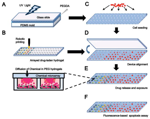
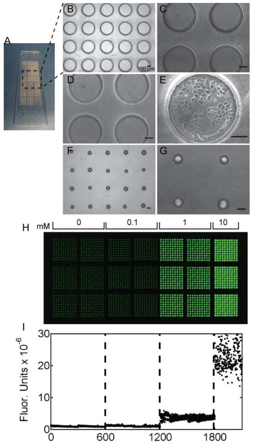
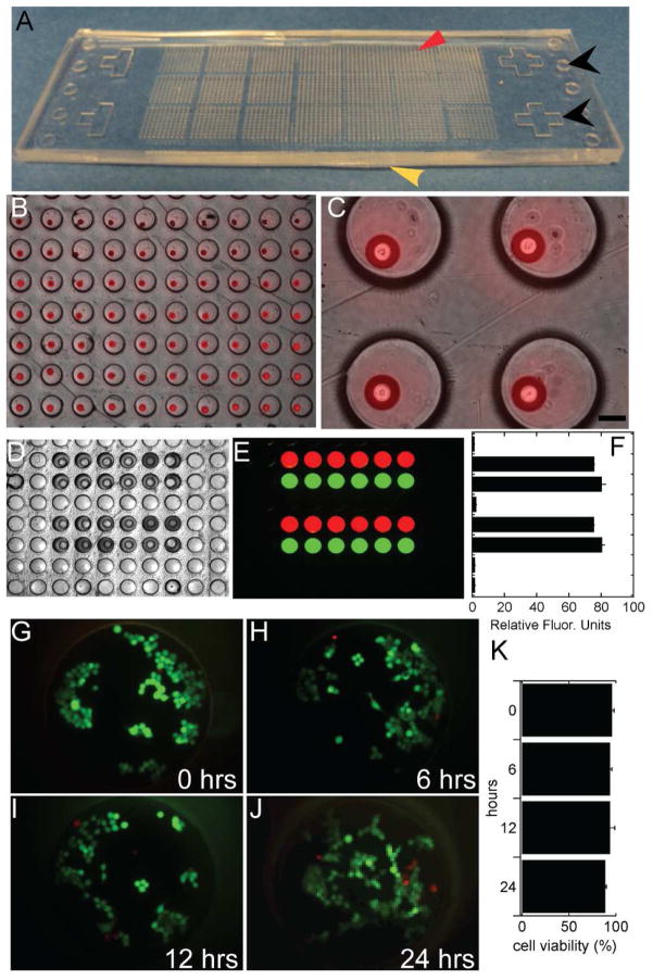
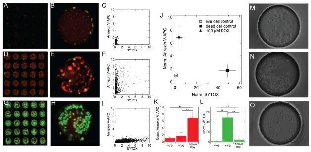
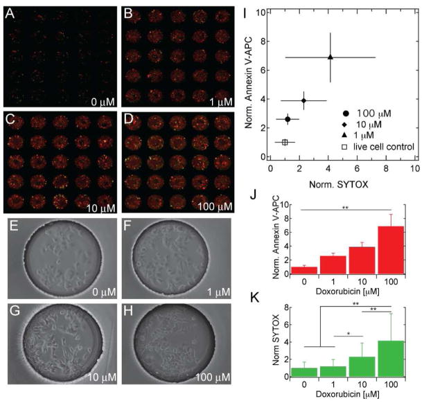
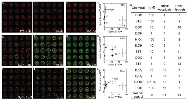
Traditional high-throughput screening (HTS) is carried out in centralized facilities that require extensive robotic liquid and plate handling equipment. This model of HTS is restrictive as such facilities are not accessible to many researchers. We have designed a simple microarray platform for cell-based screening that can be carried out at the benchtop. The device creates a microarray of 2100 individual cell-based assays in a standard microscope slide format. A microarray of chemical-laden hydrogels addresses a matching array of cell-laden microwells thus creating a microarray of sealed microscale cell cultures each with unique conditions. We demonstrate the utility of the device by screening the extent of apoptosis and necrosis in MCF-7 breast cancer cells in response to exposure to a small library of chemical compounds. From a set of screens we produced a rank order of chemicals that preferentially induce apoptosis over necrosis in MCF-7 cells. Treatment with doxorubicin induced high levels of apoptosis in comparison with staurosporine, ethanol, and hydrogen peroxide, whereas treatment with 100 μM ethanol induced minimal apoptosis with high levels of necrosis. We anticipate broad application of the device for various research and discovery applications as it is easy to use, scalable, and can be fabricated and operated with minimal peripheral equipment.


Figures

Figure 1
Figure 1
Design and fabrication of a controlled release microarray system for chemical screening. A) Micromolding of PEGDA by UV photopolymerization into arrayed microwells. B) Fabrication of a chemical-laden hydrogel microarray by robotic printing. C) Cell seeding in arrayed microwells. D) Alignment and sandwiching of arrayed chemical-laden hydrogels and cell-seeded microwells. E) Drug release and cell culturing for 6 – 24 hours. Close-up schematic shows the diffusion of chemicals from arrayed hydrogels into cell-seeded microwells. F) Analysis of apoptosis by fluorescence-based assay.
Figure 2
Figure 2
Cell-seeded microwells and arrayed chemical-laden hydrogels. A) A photograph of arrayed microwells adhered to a standard glass microscope slide. B,C) Phase contrast images of microwells (400 μm in diameter and 300 μm deep) with (D,E) seeded MCF-7 breast cancer cells. F, G) Phase contrast images of arrayed PEGDA hydrogels on a PDMS substrate. All scale bars are 100 μm. H) Fluorescent scanner image of arrayed hydrogels with varying concentrations of Rhodamine B (Ex:Em, 532:575/25; green false color). The hydrogel microarray contains 600 spots of each of 0, 0.1, and 1 mM and 300 spots of 10 mM of Rhodamine B. I) Quantification of fluorescence in each arrayed hydrogel.
Figure 3
Figure 3
Microarray device alignment and characterization. A) Photograph of a microarray device with arrayed chemical-laden hydrogels (red arrow) sandwiched on to arrayed microwells (yellow arrow). Alignment features are indicated with black arrows. B, C) Phase contrast images with overlaid fluorescent images of an aligned device (scale bar = 100 μm). D, E, F) Phase contrast and fluorescent images, and quantification of fluorescence of released chemicals (green is FITC-labeled dextran and red is Rhodamine B). G–K) MCF-7 viability in sealed microwells at 0, 6, 12 and 24 hour time points (Live/Dead staining with calcein-AM, green, and ethidium homodimer, red).
Figure 4
Figure 4
Measuring chemical-induced apoptosis. Microwell MCF-7 cell cultures exposed to (A,B) no chemicals (negative control; −ve), (D,E) 100 μM doxorubicin (DOX), and (G,H) 0.01% Triton X-100 (positive control; +ve) for 12 hours. Fluorescent images are of microwells stained with annexin V-APC (red) and SYTOX-Orange (green) (Ex:Em, 632:695/15 and 532:575/25, respectively). Pixel intensity (×10−3) due to each stain is shown in (C,F,I). The average fluorescence intensity of each control condition normalized to the negative control (−ve) is presented in (J). ANOVA analysis of the average annexin V-APC and SYTOX-Orange fluorescence for each condition are presented in (K) and (L), respectively (n ≥ 30, ** p<0.01, * p<0.05). Phase contrast images of −ve, 100 μM DOX, and +ve are shown in (M,N,O), respectively.
Figure 5
Figure 5
Increasing doxorubicin concentrations result in increased apoptosis. A–H) Fluorescent scanner and phase contrast images of microwell MCF-7 cell cultures exposed to 0, 1, 10 and 100 mM of doxorubicin. Fluorescent images show annexin V-APC (red) and SYTOX-Orange (green). I–K) Normalized annexin V-APC and SYTOX-Orange fluorescence for each doxorubicin condition, with associated ANOVA analyses (n ≥ 30, ** p<0.01, * p<0.05).
Figure 6
Figure 6
Microarrays for cell-based screening of chemical-induced apoptosis. Concentration dependent apoptosis and necrosis (as judged by annexin V-APC and SYTOX-Orange, respectively) for (A–D) staurosporine (STS), (E–H) ethanol (EtOH), and (I–L) hydrogen peroxide (H2O2). M) The rank order of all chemicals for apoptosis and necrosis as measured in the microarray device.

References

    1. Schuster D, Laggner C, Langer T. Curr Pharm Design. 2005;11(27):3545–3559. - PubMed
    1. Bleicher KH, Bohm HJ, Muller K, Alanine AI. Nat Rev Drug Discov. 2003;2(5):369–378. - PubMed
    1. Kang LF, Chung BG, Langer R, Khademhosseini A. Drug Discov Today. 2008;13(1–2):1–13. - PMC - PubMed
    1. Bailey SN, Sabatini DM, Stockwell BR. Proc Natl Acad Sci U S A. 2004;101(46):16144–16149. - PMC - PubMed
    1. Tavana H, Jovic A, Mosadegh B, Lee QY, Liu X, Luker KE, Luker GD, Weiss SJ, Takayama S. Nat Mater. 2009;8(9):736–41. - PMC - PubMed

Publication types