. 2011 Nov;22(11):1389-401.
doi: 10.1089/hum.2011.031. Epub 2011 Jun 8.

Impact of pre-existing immunity on gene transfer to nonhuman primate liver with adeno-associated virus 8 vectors

Affiliations

Impact of pre-existing immunity on gene transfer to nonhuman primate liver with adeno-associated virus 8 vectors

Lili Wang et al. Hum Gene Ther. 2011 Nov.

Abstract

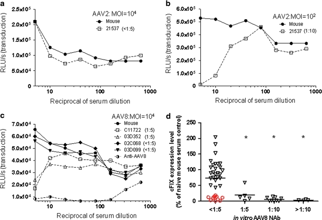
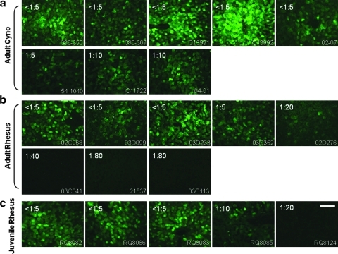
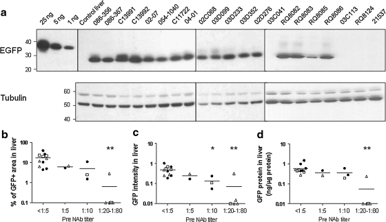
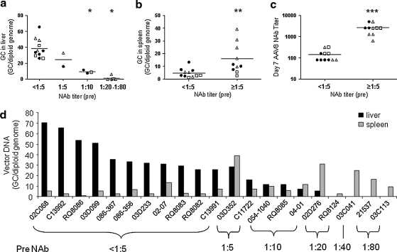
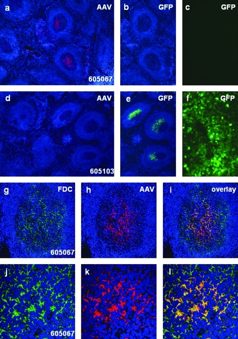
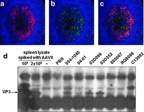
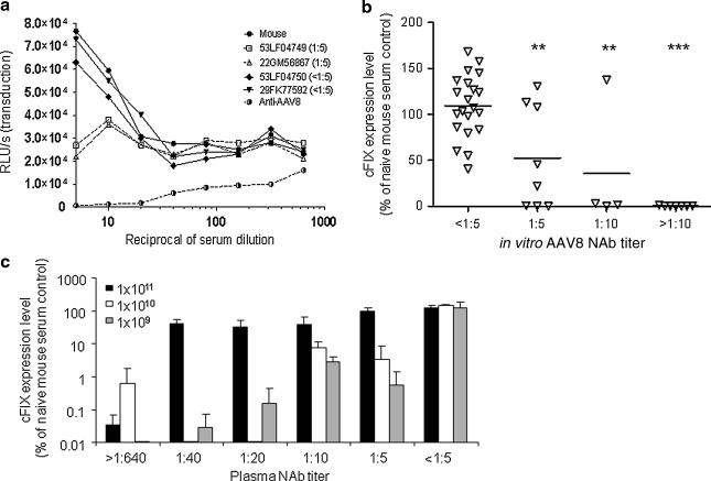
Vectors based on the primate-derived adeno-associated virus serotype 8 (AAV8) are being evaluated in preclinical and clinical models. Natural infections with related AAVs activate memory B cells that produce antibodies capable of modulating the efficacy and safety of the vector. We have evaluated the biology of AAV8 gene transfer in macaque liver, with a focus on assessing the impact of pre-existing humoral immunity. Twenty-one macaques with various levels of AAV neutralizing antibody (NAb) were injected intravenously with AAV8 vector expressing green fluorescent protein. Pre-existing antibody titers in excess of 1:10 substantially diminished hepatocyte transduction that, in the absence of NAbs, was highly efficient. Vector-specific NAb diminished liver deposition of genomes and unexpectedly increased genome distribution to the spleen. The majority of animals showed high-level and stable sequestration of vector capsid protein by follicular dendritic cells of splenic germinal centers. These studies illustrate how natural immunity to a virus that is related to a vector can impact the efficacy and potential safety of in vivo gene therapy. We propose to use the in vitro transduction inhibition assay to evaluate research subjects before gene therapy and to preclude from systemic AAV8 trials those that have titers in excess of 1:10.


Figures

FIG. 1.
FIG. 1.
Different sensitivity of in vitro neutralizing antibody (NAb) assay for AAV2 and AAV8 dictated by the in vitro transduction efficiency and a highly sensitive in vivo passive transfer (PT) assay to detect low levels of AAV8 NAb in macaque samples. Serial 2-fold dilutions of macaque and naive mouse serum were incubated with 1 × 109 genome copies (GC) (a) or 1 × 107 GC (b) of AAV2.CMV.LacZ, or with 1 × 109 GC of AAV8.CMV.LacZ (c), and transferred to preseeded 96-well plates containing 1 × 105 Huh7 cells per well. Transduction efficiency at each serum dilution was measured by determining β-galactosidase levels [relative light units (RLU) per second] 24 hr after infection. The AAV neutralization titer for each sample (indicated in parentheses) was determined as the highest serum dilution that inhibited AAV.CMV.LacZ transduction by ≥50%, compared with naive mouse serum control. To obtain a transduction signal within the dynamic range of the assay, AAV2 (MOI, 104) vector–serum mix was diluted 1:50 before transferral to the preseeded 96-well plate. (d) An in vivo PT assay to detect low levels of AAV8 NAb in macaque serum samples. Seven days after PT and vector injection, canine factor IX (cFIX) levels in plasma were measured and shown as the percentage of the cFIX level in control mice passively transferred with naive mouse serum. Open red circles (n = 10) indicate macaque samples with undetectable AAV8 NAb (<1:5) that showed inhibition by the in vivo PT assay. Mean levels for each group are shown by the horizontal lines. *p < 0.05, using an ANOVA Dunnett test when compared with the group with pre-NAb < 1:5. n = 35 (<1:5), n = 5 (1:5), n = 9 (1:10), and n = 4 (>1:10).
FIG. 2.
FIG. 2.
Transduction of macaque liver with AAV2/8 vector. EGFP expression in macaque liver 7 days after intravenous infusion of AAV2/8.TBG.EGFP at 3 × 1012 GC/kg. (a) Adult cynomolgus macaques (n = 8). (b) Adult rhesus macaques (n = 8). (c) Juvenile rhesus macaques (n = 5). Representative images of each animal are shown. Preinjection AAV8 NAb titer is indicated at the top left corner, and the animal number is shown at the bottom right corner. Scale bar: 100 μm.
FIG. 3.
FIG. 3.
Impact of pre-existing AAV8 NAb on GFP expression levels in macaque liver. (a) Western analysis of liver lysate (10 μg of protein per lane) from a control and vector-treated macaques (7 days after vector administration). Purified EGFP protein with 12-His tag was loaded at 25, 5, and 1 ng per lane as controls. (b) Quantitative morphometric analysis of the transduction efficiency based on percent transduction of area. (c) Quantitative morphometric analysis of the transduction efficiency based on relative GFP intensity. (d) Quantification of GFP protein in liver lysate by ELISA: adult cynomolgus macaques (n = 8, solid circles), adult rhesus macaques (n = 8, open triangles), juvenile rhesus macaques (n = 5, open squares). Horizontal lines show the mean levels for each group. *p < 0.05, **p < 0.01 by ANOVA Dunnett test when compared with the group with pre-NAb < 1:5.
FIG. 4.
FIG. 4.
Impact of pre-existing AAV8 NAb on vector biodistribution and day 7 NAb in macaques. (a) The amount of vector in liver (day 7) is negatively impacted by pre-existing AAV8 NAb. (b) Vector distribution to spleen is enhanced by pre-existing AAV8 NAb. (c) Dramatic increase in day 7 NAb titer in macaques with pre-existing AAV8 NAb. (d) Vector biodistribution in liver and spleen in individual animals 7 days after vector administration. Adult cynomolgus macaques (n = 8, solid circles), adult rhesus macaques (n = 8, open triangles), juvenile rhesus macaques (n = 5, open squares). Horizontal lines show the mean levels for each group. *p < 0.0001 by ANOVA Dunnett test, compared with the group with pre-NAb < 1:5. **p < 0.01, ***p < 0.0001 by Student t test.
FIG. 5.
FIG. 5.
Detection of AAV8 capsid protein or GFP protein in the germinal centers of macaque spleens. (ac) Spleen and liver from animal 605067 (pre-NAb titer, 1:10; harvested 35 days after receiving AAV8 vector). Immunostaining shows the presence of capsid protein in germinal centers of the spleen (a). GFP is undetectable in the spleen (shown on the same spleen section) (b) or in the liver (c). (df) Spleen and liver from animal 605103 (pre-NAb titer <1:5, harvested on day 35 after vector administration). No capsid protein is detectable in the spleen by immunostaining (d). The germinal centers contain GFP (e) and GFP expression is visible in the liver (f). (gi) Double immunofluorescence staining of spleen from animal 605067 with antibodies directed against follicular dendritic cells (FDCs) (g) and AAV8 capsid (h), showing colocalization (i) within germinal centers. (jl) Double staining of FDCs and capsid shown at higher magnification. Spleen images have been counterstained with DAPI (shown in blue) to visualize nuclei.
FIG. 6.
FIG. 6.
Detection of AAV8 capsid protein in macaque spleen by immunostaining and immunoprecipitation (IP). Double immunofluorescence staining of germinal centers (animal 03D352) with two different antibodies against AAV8: (a) AAV8-immunized rabbit serum, (b) AAV8 capsid-specific monoclonal antibody ADK8, and (c) overlay of both stains. Sections have been stained with DAPI (blue) to show nuclei. (d) Detection of VP3 protein in some nonhuman primate (NHP) spleen lysates by IP and Western blot. IP was performed with anti-AAV8 rabbit serum to pull down AAV capsid proteins from 0.5 mg (animals 605067 and RQ8086) or 1.5 mg (the remaining five animals) of spleen lysate. A negative spleen sample was spiked in with 1 × 109 or 2 × 108 GC of AAV8 to serve as positive controls. The capsid proteins were further detected by Western blot, using the monoclonal B1 antibody.
FIG. 7.
FIG. 7.
In vitro and in vivo assays to detect AAV8 NAb in human plasma samples. (a) In vitro NAb assay. The AAV8 NAb titer for each sample is indicated in parentheses. (b) Correlation of in vitro NAb titer and the outcome of in vivo passive transfer (n = 40 samples, vector dose = 1 × 109 GC). (c) Effect of vector dose on passive transfer. Human plasma samples with various AAV8 NAb titers or naive mouse serum was passively transferred to C57BL/6 mice (200 μl/mouse) via the tail vein. Two hours later, mice were intravenously injected with 1 × 109, 1 × 1010, or 1 × 1011 GC of AAV2/8.LSP.cFIX-W (n = 3 for each dose). Seven days after vector injection, cFIX levels in plasma were measured by ELISA and are shown as the percentage of the cFIX level in control mice passively transferred with naive mouse serum and receiving the same vector dose. Means and SD are shown. **p < 0.01, ***p < 0.001 by ANOVA Dunnett test, compared with the group with pre-NAb < 1:5.

References

    1. Bainbridge J.W. Smith A.J. Barker S.S., et al. Effect of gene therapy on visual function in Leber's congenital amaurosis. N. Engl. J. Med. 2008;358:2231–2239. - PubMed
    1. Barker S.E. Broderick C.A. Robbie S.J., et al. Subretinal delivery of adeno-associated virus serotype 2 results in minimal immune responses that allow repeat vector administration in immunocompetent mice. J. Gene Med. 2009;11:486–497. - PMC - PubMed
    1. Bell P. Moscioni A.D. McCarter R.J., et al. Analysis of tumors arising in male B6C3F1 mice with and without AAV vector delivery to liver. Mol. Ther. 2006;14:34–44. - PubMed
    1. Boutin S. Monteilhet V. Veron P., et al. Prevalence of serum IgG and neutralizing factors against adeno-associated virus (AAV) types 1, 2, 5, 6, 8, and 9 in the healthy population: Implications for gene therapy using AAV vectors. Hum. Gene Ther. 2010;21:704–712. - PubMed
    1. Brantly M.L. Chulay J.D. Wang L., et al. Sustained transgene expression despite T lymphocyte responses in a clinical trial of rAAV1-AAT gene therapy. Proc. Natl. Acad. Sci. U.S.A. 2009;106:16363–16368. - PMC - PubMed

Publication types

Substances

LinkOut - more resources