. 2011 Jun;153(5):634-43.
doi: 10.1111/j.1365-2141.2011.08679.x. Epub 2011 Apr 8.

Dyskeratosis congenita and the DNA damage response

Affiliations
Free PMC article

Dyskeratosis congenita and the DNA damage response

Michael Kirwan et al. Br J Haematol. 2011 Jun.
Free PMC article

Abstract

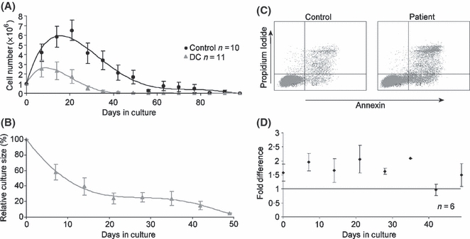
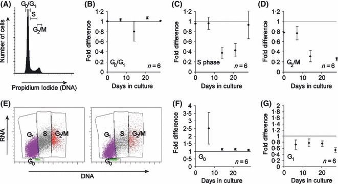
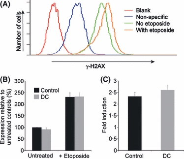
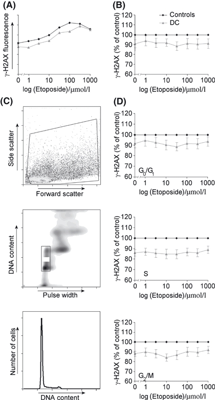
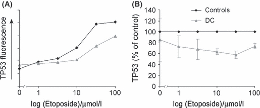
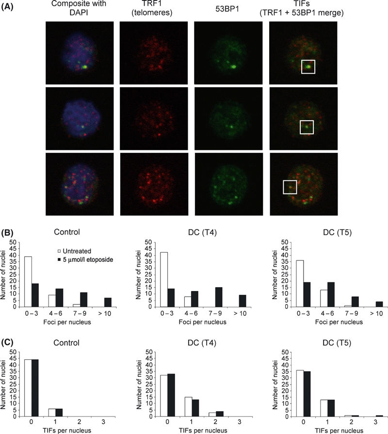
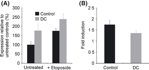
Dyskeratosis congenita (DC) is a heterogeneous bone marrow failure disorder with known mutations in components of telomerase and telomere shelterin. Recent work in a mouse model with a dyskerin mutation has implicated an increased DNA damage response as part of the cellular pathology, while mouse models with Terc and Tert mutations displayed a normal response. To clarify how these contradictory results might apply to DC pathology in humans, we studied the cellular phenotype in primary cells from DC patients of several genetic subtypes, focussing on T lymphocytes to remain close to the haematopoietic system. We observed novel cell cycle abnormalities in conjunction with impaired growth and an increase in apoptosis. Using flow cytometry and confocal microscopy we examined induction of the DNA damage proteins γ-H2AX and 53BP1 and the cell cycle protein TP53 (p53). We found an increase in damage foci at telomeres in lymphocytes and an increase in the basal level of DNA damage in fibroblasts, but crucially no increased response to DNA damaging agents in either cell type. As the response to induced DNA damage was normal and levels of global DNA damage were inconsistent between cell types, DNA damage may contribute differently to the pathology in different tissues.


Figures

Fig 1
Fig 1
T lymphocytes from DC patients show impaired growth and increased apoptosis. (A) Cell numbers counted every 7 d in T lymphocyte cultures. 1 × 10⁶ cells were plated at day 0 and stimulated with PHA for 48 h. Cells were subsequently cultured in interleukin-2 supplemented medium. (B) DC lymphocyte cell numbers expressed as a percentage of control cell numbers (control n = 10) show an increasing growth deficit over the culture period. (C) Propidium iodide and annexin V co-staining of lymphocytes as measured by flow cytometry using sample D1 as an example. On the left is the control sample and on the right the patient sample. (D) A higher proportion of DC lymphocytes (around twofold higher than controls) were apoptotic over 49 d in culture. Error bars represent the standard error from six patient sample measurements (four TERC, two DKC1) against six paired controls.
Fig 2
Fig 2
Cell cycle abnormalities in DC lymphocytes after mitogenic stimulation (A) Nuclear staining of lymphocytes with propidium iodide as measured by flow cytometry. The phases of the cell cycle are marked. (B) More DC lymphocytes are in the G0/G1 phase compared to controls. (C) Fewer DC lymphocytes are in the S phase compared to controls. (D) Fewer DC lymphocytes are in the G2/M phase compared to controls. (E) Co-staining of lymphocyte DNA with Hoescht 33342 and RNA with pyronin Y as measured by flow cytometry using sample D1 as an example. On the left is the control and on the right the patient sample. The phases of the cell cycle are marked. (F) More DC lymphocytes are in the G0 phase compared to controls. (G) Fewer DC lymphocytes are in the G1 phase compared to controls. All graphs express fold difference to control samples. Error bars represent the standard error from six patient sample measurements (four TERC, two DKC1) against six paired controls.
Fig 3
Fig 3
The DNA damage response is not up-regulated in T lymphocytes from our DC cohort. (A) DNA damage associated histone protein γ-H2AX expression as measured by flow cytometry. Fluorescence is measured in the absence of antibodies to determine levels of auto-fluorescence (blank). Fluorescence in the presence of fluorescently-labelled secondary antibody only is regarded as non-specific and is subtracted from all subsequent measurements. Fluorescence in the presence of primary (anti-γ-H2AX) antibody plus secondary is considered specific due to the presence of γ-H2AX. Incubation with etoposide causes a net increase in median fluorescence due to increased activation of H2AX to γ-H2AX. (B) Relative expression of γ-H2AX in DC lymphocytes compared to controls both with and without 350 μmol/l etoposide exposure shows no significant difference. (C) Fold induction of γ-H2AX expression in DC lymphocytes is not significantly different to controls. Error bars represent the standard error from six DC samples (three TERC, two DKC1, one TERT) against six paired controls.
Fig 4
Fig 4
γ-H2AX expression in T lymphocytes assayed in detail across a range of etoposide doses and cell cycle populations. (A) Example of a γ-H2AX dose-response curve for etoposide exposure showing a control sample and a DC sample in the same assay. (B) γ-H2AX expression of nine DC samples (five TERC, three DKC1 and one TERT) normalized to seven paired controls. (C) After acquisition of whole cells (upper panel) DAPI staining of the nuclei allows exclusion of doublets (middle panel) and cells in different phases of the cell cycle can be specifically gated (lower panel). (D) Dose-response curves showing γ-H2AX expression in different phases of the cell cycle.
Fig 5
Fig 5
Expression of TP53 in lymphocytes after 6 h of etoposide exposure across a range of concentrations. (A) Example showing that T lymphocytes show a dose-response in TP53 expression after etoposide treatment. (B) There is no increase in TP53 expression in DC cells (one TERC, one TERT) compared to two paired controls.
Fig 6
Fig 6
Confocal microscopy showing DNA damage foci in T lymphocytes. (A) Three example images showing nuclei stained with DAPI and labelled for TRF1 telomeric protein, 53BP1 DNA damage protein and a merged image showing 53BP1 colocalizing with telomeres at telomere induced damage foci (TIFs). (B) The number of 53BP1 DNA damage foci per nucleus is similar in a control sample and in two DC samples. (C) There is a small but significant increase in the number of TIFs in the DC lymphocytes. In all cases 50 nuclei were counted.
Fig 7
Fig 7
The basal level of DNA damage is increased in DC fibroblasts but the DNA damage response is normal. (A) Relative expression of γ-H2AX in fibroblasts compared to controls both with and without 350 μmol/l etoposide exposure shows a significantly higher level of expression in DC cells. (B) Fold induction of γ-H2AX expression in DC fibroblasts is not significantly different to controls. Error bars represent the standard error from three DC samples (three DKC1) against two controls.

References

    1. d'Adda di Fagagna F, Reaper PM, Clay-Farrace L, Fiegler H, Carr P, Von Zglinicki T, Saretzki G, Carter NP, Jackson SP. A DNA damage checkpoint response in telomere-initiated senescence. Nature. 2003;426:194–198. - PubMed
    1. Alter BP, Baerlocher GM, Savage SA, Chanock SJ, Weksler BB, Willner JP, Peters JA, Giri N, Lansdorp PM. Very short telomere length by flow fluorescence in situ hybridization identifies patients with dyskeratosis congenita. Blood. 2007;110:1439–1447. - PMC - PubMed
    1. Armanios M, Chen JL, Chang YP, Brodsky RA, Hawkins A, Griffin CA, Eshleman JR, Cohen AR, Chakravarti A, Hamosh A, Greider CW. Haploinsufficiency of telomerase reverse transcriptase leads to anticipation in autosomal dominant dyskeratosis congenita. Proceedings of the National Academy of Sciences of the United States of America. 2005;102:15960–15964. - PMC - PubMed
    1. Auerbach AD. Fanconi anemia diagnosis and the diepoxybutane (DEB) test. Experimental Hematology. 1993;21:731–733. - PubMed
    1. Bellodi C, Kopmar N, Ruggero D. Deregulation of oncogene-induced senescence and p53 translational control in X-linked dyskeratosis congenita. EMBO Journal. 2010;29:1865–1876. - PMC - PubMed

Publication types

MeSH terms

Substances