. 2011 Nov;15(11):2273-83.
doi: 10.1111/j.1582-4934.2011.01327.x.

Npp1 promotes atherosclerosis in ApoE knockout mice

Affiliations

Npp1 promotes atherosclerosis in ApoE knockout mice

Yvonne Nitschke et al. J Cell Mol Med. 2011 Nov.

Abstract

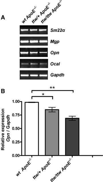
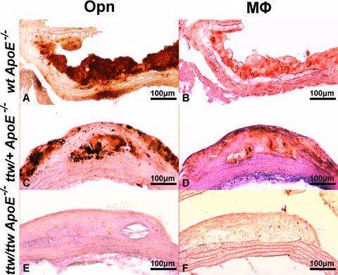
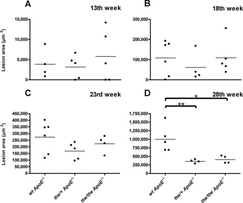
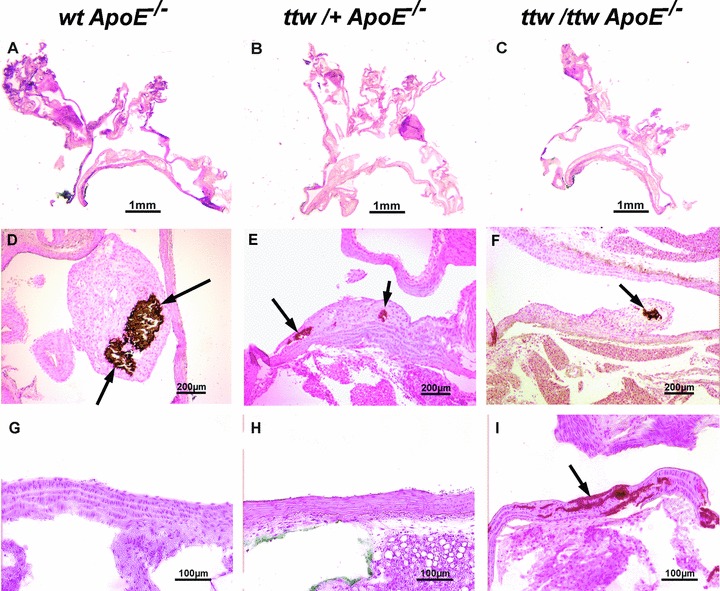
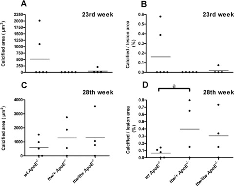
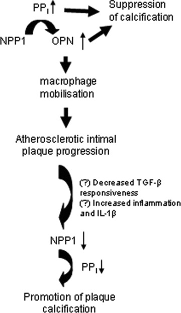
Ecto-nucleotide pyrophosphatase/phosphodiesterase 1 (NPP1) generates inorganic pyrophosphate (PP(i)), a physiologic inhibitor of hydroxyapatite deposition. In a previous study, we found NPP1 expression to be inversely correlated with the degree of atherosclerotic plaque calcification. Moreover, function-impairing mutations of ENPP1, the gene encoding for NPP1, are associated with severe, artery tunica media calcification and myointimal hyperplasia with infantile onset in human beings. NPP1 and PP(i) have the potential to modulate atherogenesis by regulating arterial smooth muscle cell (SMC) differentiation and function, including increase of pro-atherogenic osteopontin (OPN) expression. Hence, this study tested the hypothesis that NPP1 deficiency modulates both atherogenesis and atherosclerotic intimal plaque calcification. Npp1/ApoE double deficient mice were generated by crossing mice bearing the ttw allele of Enpp1 (that encodes a truncation mutation) with ApoE null mice and fed with high-fat/high-cholesterol atherogenic diet. Atherosclerotic lesion area and calcification were examined at 13, 18, 23 and 28 weeks of age. The aortic SMCs isolated from both ttw/ttw ApoE(-/-) and ttw/+ ApoE(-/-) mice demonstrated decreased Opn expression. The 28-week-old ttw/ttw ApoE(-/-) and ttw/+ ApoE(-/-) had significantly smaller atherosclerotic lesions compared with wild-type congenic ApoE(-/-) mice. Only ttw/ttw but not ttw/+ mice developed artery media calcification. Furthermore in ttw/+ mice, there was a tendency towards increased plaque calcification compared to ApoE(-/-) mice without Npp1 deficiency. We conclude that Npp1 promotes atherosclerosis, potentially mediated by Opn expression in ApoE knockout mice.


Figures

Fig 1
Fig 1
RT-PCR analysis for chondro-osseous gene expression in cultured aortic SMC preparations from wt ApoE−/−, ttw/+ ApoE−/− and ttw/ttw ApoE−/− mice. (A) Representative RT-PCR of one of four individual animals of each genotype is shown. (B) Relative expression of Opn corrected for housekeeping gene Gapdh. Aortic SMC from 6-week-old mice were harvested as described in ‘Material and methods’. The mRNAs were studied by RT-PCR in aliquots of 3 × 10⁴ cells cultured for eight days as described in ‘Material and methods’. Values represent the data mean ± S.E.M. One-way anova was used to determine significant differences with Tukey’s post-hoc analysis. *P < 0.05, **P < 0.001.
Fig 2
Fig 2
Representative stainings of Opn and macrophages in atherosclerotic lesions of 28-week-old mice. Opn (brown, A, C, E) and macrophages (brown, B, D, F) immunohistochemistry in wt ApoE−/− (A, B), ttw/+ ApoE−/− (C, D) and ttw/ttw ApoE−/− (E, F) mice fed a high-fat/high-cholesterol diet.
Fig 4
Fig 4
Mean atherosclerosis lesion area in individual mice sorted by genotype and age. Mean lesion area in μm2 was calculated in 13 (A), 18 (B), 23 (C) and 28-week-old mice (D). Bars represent the data mean. One-way anova was used to determine significant differences with Tukey’s post-hoc analysis. *P < 0.05 versus wt ApoE−/− mice, **P < 0.01 versus wt ApoE−/− mice.
Fig 3
Fig 3
Representative aortic root sections of 28-week-old mice. wt ApoE−/− (A, D, G), ttw/+ ApoE−/− (B, E, H) and ttw/ttw ApoE−/− (C, F, I) mice fed a high-fat/high-cholesterol diet. Haematoxylin and eosin stained sections (A–C) of the whole aortic arch. Von Kossa staining for calcifications (brown, arrows) of atherosclerostic lesions (D–F) and the aortic wall (G–I).
Fig 5
Fig 5
Calcified areas in individual mice, sorted by genotype and age. Mean calcified area in μm2 (A and C) and calcified area expressed as percentage of total lesion area (B and D) determined in 23- (A and B) and 28- (C and D) week-old mice. Bars represent the data mean. One-way anova was used to determine significant differences with Tukey’s post-hoc analysis. aP < 0.1 versus wt ApoE−/− mice.
Fig 6
Fig 6
Model for modulation of NPP1 expression and role of NPP1 in atherogenesis and suppression of intimal plaque calcification based on results of promotion of atherosclerotic lesion development but suppression of intimal plaque calcification. This model is based on the results of a previous study that showed regulated expression of NPP1 and OPN in human atherosclerotic lesions [29], and is based on the results of the current study in ApoE knockout mice with partial and severe deficiency of Npp1 in ttw/+ and ttw/ttw mice. NPP1 generates PPi and thereby promotes OPN expression that promotes progression of atherogenesis. However, a shift within lesions to decreased TGF-β responsiveness [45] and increased inflammation including IL-1β expression [43, 44] could account for the previously described drop NPP1 expression in atherosclerotic lesion cells including SMCs. This effect, linked with decreased PPi generation and inhibition of OPN expression, promotes plaque calcification. We observe increased atherosclerotic plaque calcification in Npp1-/ApoE-deficient lesions in vivo in this study.

References

    1. Watson KE, Demer LL. The atherosclerosis-calcification link. Curr Opin Lipidol. 1996;7:101–4. - PubMed
    1. Wilson PW, Kauppila LI, O’Donnell CJ, et al. Abdominal aortic calcific deposits are an important predictor of vascular morbidity and mortality. Circulation. 2001;103:1529–34. - PubMed
    1. Wayhs R, Zelinger A, Raggi P. High coronary artery calcium scores pose an extremely elevated risk for hard events. J Am Coll Cardiol. 2002;39:225–30. - PubMed
    1. Speer MY, Li X, Hiremath PG, et al. Runx2/Cbfa1, but not loss of myocardin, is required for smooth muscle cell lineage reprogramming toward osteochondrogenesis. J Cell Biochem. 2010;110:935–47. - PMC - PubMed
    1. Speer MY, Yang HY, Brabb T, et al. Smooth muscle cells give rise to osteochondrogenic precursors and chondrocytes in calcifying arteries. Circ Res. 2009;104:733–41. - PMC - PubMed

Publication types

MeSH terms