Parasitostatic effect of maslinic acid. I. Growth arrest of Plasmodium falciparum intraerythrocytic stages
- PMID: 21477369
- PMCID: PMC3087696
- DOI: 10.1186/1475-2875-10-82
Parasitostatic effect of maslinic acid. I. Growth arrest of Plasmodium falciparum intraerythrocytic stages
Abstract
Background: Natural products have played an important role as leads for the development of new drugs against malaria. Recent studies have shown that maslinic acid (MA), a natural triterpene obtained from olive pomace, which displays multiple biological and antimicrobial activities, also exerts inhibitory effects on the development of some Apicomplexan, including Eimeria, Toxoplasma and Neospora. To ascertain if MA displays anti-malarial activity, the main objective of this study was to asses the effect of MA on Plasmodium falciparum-infected erythrocytes in vitro.
Methods: Synchronized P. falciparum-infected erythrocyte cultures were incubated under different conditions with MA, and compared to chloroquine and atovaquone treated cultures. The effects on parasite growth were determined by monitoring the parasitaemia and the accumulation of the different infective stages visualized in thin blood smears.
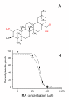
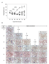
Results: MA inhibits the growth of P. falciparum Dd2 and 3D7 strains in infected erythrocytes in, dose-dependent manner, leading to the accumulation of immature forms at IC50 concentrations, while higher doses produced non-viable parasite cells. MA-treated infected-erythrocyte cultures were compared to those treated with chloroquine or atovaquone, showing significant differences in the pattern of accumulation of parasitic stages. Transient MA treatment at different parasite stages showed that the compound targeted intra-erythrocytic processes from early-ring to schizont stage. These results indicate that MA has a parasitostatic effect, which does not inactivate permanently P. falciparum, as the removal of the compound allowed the infection to continue
Conclusions: MA displays anti-malarial activity at multiple intraerythrocytic stages of the parasite and, depending on the dose and incubation time, behaves as a plasmodial parasitostatic compound. This novel parasitostatic effect appears to be unrelated to previous mechanisms proposed for current anti-malarial drugs, and may be relevant to uncover new prospective plasmodial targets and opens novel possibilities of therapies associated to host immune response.
Figures


Similar articles
-
Parasitostatic effect of maslinic acid. II. Survival increase and immune protection in lethal Plasmodium yoelii-infected mice.Malar J. 2011 Apr 25;10:103. doi: 10.1186/1475-2875-10-103. Malar J. 2011. PMID: 21518429 Free PMC article.
-
In vitro and in vivo properties of ellagic acid in malaria treatment.Antimicrob Agents Chemother. 2009 Mar;53(3):1100-6. doi: 10.1128/AAC.01175-08. Epub 2008 Nov 17. Antimicrob Agents Chemother. 2009. PMID: 19015354 Free PMC article.
-
Drug-induced death of the asexual blood stages of Plasmodium falciparum occurs without typical signs of apoptosis.Microbes Infect. 2006 May;8(6):1560-8. doi: 10.1016/j.micinf.2006.01.016. Epub 2006 Apr 18. Microbes Infect. 2006. PMID: 16702009
-
[In vitro cultivation of Plasmodium falciparum. Applications and limits.- Methodology].Med Trop (Mars). 1982 Jul-Aug;42(4):437-62. Med Trop (Mars). 1982. PMID: 6755144 Review. French.
-
Atovaquone/proguanil: a review of its use for the prophylaxis of Plasmodium falciparum malaria.Drugs. 2003;63(6):597-623. doi: 10.2165/00003495-200363060-00006. Drugs. 2003. PMID: 12656656 Review.
Cited by
-
Parasitostatic effect of maslinic acid. II. Survival increase and immune protection in lethal Plasmodium yoelii-infected mice.Malar J. 2011 Apr 25;10:103. doi: 10.1186/1475-2875-10-103. Malar J. 2011. PMID: 21518429 Free PMC article.
-
Analogs of natural aminoacyl-tRNA synthetase inhibitors clear malaria in vivo.Proc Natl Acad Sci U S A. 2014 Dec 23;111(51):E5508-17. doi: 10.1073/pnas.1405994111. Epub 2014 Dec 8. Proc Natl Acad Sci U S A. 2014. PMID: 25489076 Free PMC article.
-
Evaluation of in vivo anti-malarial potential of omidun obtained from fermented maize in Ibadan, Nigeria.Malar J. 2020 Nov 19;19(1):414. doi: 10.1186/s12936-020-03486-0. Malar J. 2020. PMID: 33213477 Free PMC article.
-
In vitro inhibitory effects of plant-derived by-products against Cryptosporidium parvum.Parasite. 2016;23:41. doi: 10.1051/parasite/2016050. Epub 2016 Sep 14. Parasite. 2016. PMID: 27627637 Free PMC article.
-
Investigation of Antiparasitic Activity of 10 European Tree Bark Extracts on Toxoplasma gondii and Bioguided Identification of Triterpenes in Alnus glutinosa Barks.Antimicrob Agents Chemother. 2022 Jan 18;66(1):e0109821. doi: 10.1128/AAC.01098-21. Epub 2021 Oct 11. Antimicrob Agents Chemother. 2022. PMID: 34633849 Free PMC article.
References
Publication types
MeSH terms
Substances
LinkOut - more resources
Full Text Sources

