Clinical Trial
. 2011 Dec;38(6):1361-7.
doi: 10.1183/09031936.00197710. Epub 2011 Apr 8.

An open-label trial of rituximab therapy in pulmonary alveolar proteinosis

Affiliations
Clinical Trial

An open-label trial of rituximab therapy in pulmonary alveolar proteinosis

M S Kavuru et al. Eur Respir J. 2011 Dec.

Abstract

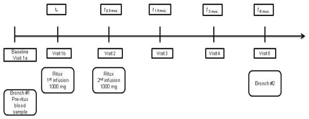
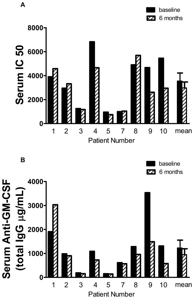
Rituximab, a monoclonal antibody directed against the B-lymphocyte antigen CD20, has shown promise in several autoimmune disorders. Pulmonary alveolar proteinosis (PAP) is an autoimmune disorder characterised by autoantibodies to granulocyte-macrophage colony-stimulating factor (GM-CSF). An open-label, proof-of-concept phase II clinical trial was conducted in 10 PAP patients. The intervention consisted of two intravenous infusions of rituximab (1,000 mg) 15 days apart. Bronchoalveolar lavage (BAL) fluid and peripheral blood samples were collected. The primary outcome was improvement in arterial blood oxygenation. Both arterial oxygen tension and alveolar-arterial oxygen tension difference in room air improved in seven out of the nine patients completing the study. Lung function and high-resolution computed tomography scans, which were secondary outcomes, also improved. Peripheral blood CD19+ B-lymphocytes decreased from mean ± sem 15 ± 2% to <0.05% (n = 10) 15 days post-therapy. This decrease persisted for 3 months in all patients; at 6 months, CD19+ B-cells were detected in four out of seven patients (5 ± 2%). Total anti-GM-CSF immunoglobulin (Ig)G levels from baseline to 6 months were decreased in BAL fluids (n = 8) but unchanged in sera (n = 9). In this PAP cohort: 1) rituximab was well-tolerated and effectively ameliorated lung disease; and 2) reduction in anti-GM-CSF IgG levels in the lung correlated with disease changes, suggesting that disease pathogenesis is related to autoantibody levels in the target organ.

Trial registration: ClinicalTrials.gov NCT00552461.


Figures

Figure 1
Figure 1
Schematic of study visits for rituximab trial
Figure 2
Figure 2. Lung function parameters are improved with rituximab treatment
(A) PaO2 was measured at baseline (before rituximab), 3 and 6 months post treatment in all patients except #1 where a 3 month sample was not obtained and #6 who refused clinical follow-up after visit 3. (B) A-a gradient was measured pre- and 3 and 6 months post-rituximab. (C) High resolution computed tomography (HRCT) demonstrated improvement with rituximab therapy. The effect of rituximab therapy on the severity of pulmonary alveolar proteinosis (PAP) lung disease was measured by the zonal HRCT score [19] as described in the supplement. (D) HRCT scan of a representative mid zone of patient #8 pre- and post-rituximab therapy.
Figure 3
Figure 3. Rituximab therapy depletes B cells
CD19+ cells were determined by flow cytometric analysis of blood samples at each study visit.
Figure 4
Figure 4
(A–B) Serum GM-CSF neutralizing capacity and total anti-GM-CSF immunoglobulin G (IgG) was not altered with rituximab therapy. Serum samples were obtained pre- and post-rituximab therapy. GM-CSF neutralizing activity was measured by the TF-1 assay as previously described [10] and anti-GM-CSF antibody was determined by a luminex based assay as described previously.[22] (C–D) Bronchial alveolar lavage (BAL) fluid GM-CSF neutralizing capacity and total anti-GM-CSF IgG was decreased with rituximab therapy. BAL fluid was obtained pre- and post-therapy and antibody levels were measured as described for serum.
Figure 4
Figure 4
(A–B) Serum GM-CSF neutralizing capacity and total anti-GM-CSF immunoglobulin G (IgG) was not altered with rituximab therapy. Serum samples were obtained pre- and post-rituximab therapy. GM-CSF neutralizing activity was measured by the TF-1 assay as previously described [10] and anti-GM-CSF antibody was determined by a luminex based assay as described previously.[22] (C–D) Bronchial alveolar lavage (BAL) fluid GM-CSF neutralizing capacity and total anti-GM-CSF IgG was decreased with rituximab therapy. BAL fluid was obtained pre- and post-therapy and antibody levels were measured as described for serum.

References

    1. Grillo-Lopez AJ, White CA, Varns C, Shen D, Wei A, McClure A, Dallaire BK. Overview of the clinical development of rituximab: first monoclonal antibody approved for the treatment of lymphoma. Semin Oncol. 1999;26:66–73. - PubMed
    1. Anolik J, Sanz I, Looney RJ. B cell depletion therapy in systemic lupus erythematosus. Curr Rheumatol Rep. 2003;5:350–356. - PubMed
    1. Edwards JC, Szczepanski L, Szechinski J, Filipowicz-Sosnowska A, Emery P, Close DR, Stevens RM, Shaw T. Efficacy of B-cell-targeted therapy with rituximab in patients with rheumatoid arthritis. N Engl J Med. 2004;350:2572–2581. - PubMed
    1. Stone JH, Merkel PA, Spiera R, Seo P, Langford CA, Hoffman GS, Kallenberg CGM, StClair EW, Turkiewicz A, Tchao NK, et al. Rituximab versus Cyclophosphamide for ANCA-Associated Vasculitis. N Engl J Med. 2010;363:221–232. - PMC - PubMed
    1. Cambridge G, Leandro MJ, Edwards JC, Ehrenstein MR, Salden M, Bodman-Smith M, Webster AD. Serologic changes following B lymphocyte depletion therapy for rheumatoid arthritis. Arthritis Rheum. 2003;48:2146–2154. - PubMed

Publication types

MeSH terms

Associated data