. 2011 May 1;25(9):907-16.
doi: 10.1101/gad.2038411. Epub 2011 Apr 8.

Endogenous Myc maintains the tumor microenvironment

Affiliations

Endogenous Myc maintains the tumor microenvironment

Nicole M Sodir et al. Genes Dev. .

Abstract

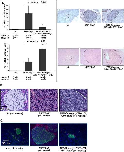
The ubiquitous deregulation of Myc in human cancers makes it an intriguing therapeutic target, a notion supported by recent studies in Ras-driven lung tumors showing that inhibiting endogenous Myc triggers ubiquitous tumor regression. However, neither the therapeutic mechanism nor the applicability of Myc inhibition to other tumor types driven by other oncogenic mechanisms is established. Here, we show that inhibition of endogenous Myc also triggers ubiquitous regression of tumors in a simian virus 40 (SV40)-driven pancreatic islet tumor model. Such regression is presaged by collapse of the tumor microenvironment and involution of tumor vasculature. Hence, in addition to its diverse intracellular roles, endogenous Myc serves an essential and nonredundant role in coupling diverse intracellular oncogenic pathways to the tumor microenvironment, further bolstering its credentials as a pharmacological target.


Figures

Figure 1.
Figure 1.
Endogenous Myc function is required for progression and maintenance of RIP1-Tag2-induced tumors. (A) Hematoxylin and eosin (H&E) staining of pancreata sections from 14-wk-old nontransgenic controls (ctr), 7-wk-old RIP1-Tag2, 14-wk-old RIP1-Tag2, and 14-wk-old TRE-Omomyc;CMVrtTA;RIP1-Tag2 mice treated with doxycycline for 7 wk prior to sacrifice. Yellow broken lines delineate islet/tumor borders. Ages of mice at sacrifice are shown in brackets. (B) H&E staining of pancreata sections from 14-wk-old nontransgenic controls (ctr), 11-wk-old RIP1-Tag2, 14-wk-old untreated TRE-Omomyc;CMVrtTA;RIP1-Tag2 mice, and 14-wk-old TRE-Omomyc;CMVrtTA;RIP1-Tag2 mice treated with doxycycline for 3 wk prior to sacrifice. Yellow broken lines delineate islet borders. Ages of mice at sacrifice are shown in brackets (see also Supplemental Figs. 1–4).
Figure 2.
Figure 2.
Inhibition of endogenous Myc function decreases proliferation and increases apoptosis in RIP1-Tag2 pancreatic β cells but does not suppress SV40 T/t-induced cellular transformation or disrupt α-cell disposition. (A) Immunohistochemical quantitation of Ki67-positive (top) and TUNEL-positive (bottom) cells in islets from 14-wk-old nontransgenic control (ctr), RIP1-Tag2, and TRE-Omomyc;CMVrtTA;RIP1-Tag2 mice treated with doxycycline for 1 wk prior to sacrifice. Data are represented as mean + SD. Representative images of islet/β-cell tumors used in this immunohistochemical quantitation are shown on the right (see also Supplemental Fig. 5). (B) Higher-magnification images of H&E-stained islets isolated from 14-wk-old nontransgenic control (ctr), RIP1-Tag2, and TRE-Omomyc;CMVrtTA;RIP1-Tag2 mice treated with doxycycline for 3 wk prior to sacrifice showing aberrant transformed morphology of β cells in RIP1-Tag2 and TRE-Omomyc;CMVrtTA;RIP1-Tag2 β-cell tumors. (C) Immunohistochemical staining of glucagon-expressing α cells (in red) and insulin-expressing β cells (in green) in islets from 14-wk-old nontransgenic control (ctr), RIP1-Tag2, and TRE-Omomyc;CMVrtTA;RIP1-Tag2 mice treated with doxycycline for 3 wk prior to sacrifice. Nuclear Hoechst counterstaining is shown in blue.
Figure 3.
Figure 3.
Endogenous Myc is required for maintenance of RIP1-Tag2 tumor vasculature. (A) Immunostaining with hypoxyprobe-1 antibody (darker areas indicated by arrowheads indicate hypoxic regions superimposed on Fast Red Nuclear counterstain) in sections of pancreata harvested from 14-wk-old nontransgenic control (ctr) mice (left), RIP1-Tag2 mice (middle), and TRE-Omomyc;CMVrtTA;RIP1-Tag2 mice (right) treated with doxycycline for 1 wk prior to sacrifice. Broken lines delineate tumor borders. (B) Immunostaining for the endothelial cell marker Meca32 (in red) and apoptosis (TUNEL in green) of sections from pancreata harvested from the same mice as in A. Insets iiii show representative higher-magnification images of TUNEL-positive apoptotic endothelial cells (see also Supplemental Fig. 6).
Figure 4.
Figure 4.
Relative kinetics of onset of endothelial versus β-cell apoptosis in RIP1-Tag2 tumors after Myc inhibition shows that vascular apoptosis precedes that of tumor cells. Quantification of active Caspase 3-positive (Casp 3*) cells in the endothelial versus β-cell compartment of TRE-Omomyc;CMVrtTA;RIP1-Tag2 tumors treated for 0, 3, or 7 d with doxycycline. Data are represented as mean + SD.
Figure 5.
Figure 5.
Endogenous Myc is required for binding of VEGF–VEGFR interactions in RIP1-Tag2-induced tumors. Immunohistochemical analysis of VEGF:VEGF-R2 conjugates (in red) and Meca32 (in green) in sections of pancreata harvested from 14-wk-old nontransgenic control (ctr), RIP1-Tag2, and TRE-Omomyc;CMVrtTA;RIP1-Tag2 mice treated with doxycycline for 1 wk prior to sacrifice. Nuclear Hoechst counterstaining (in blue) of adjacent sections is shown in the top panels. Broken yellow lines mark tumor borders.
Figure 6.
Figure 6.
Endogenous Myc is required for retention of inflammatory cells by RIP1-Tag2 tumors. Immunohistochemical analysis of neutrophils (top panels, in pink over blue Hoechst counterstain) and macrophages (bottom panels, in green over blue Hoechst counterstain) in RIP1-Tag2 and TRE-Omomyc;CMVrtTA;RIP1-Tag2 mice treated at 13 wk of age for 3 d with doxycycline prior to sacrifice. Broken yellow lines delineate the islet borders.
Figure 7.
Figure 7.
Myc inhibition rapidly modulates proinflammatory and anti-inflammatory genes. Comparative gene expression microarray analysis of cytokine, chemokine-encoding, and inflammation-related genes from laser-captured islets from 13-wk-old TRE-Omomyc;CMVrtTA;RIP1-Tag2 mice treated for 2 d with doxycycline versus untreated (sucrose only). The ratio of treated/untreated expression is represented by blue bars for down-regulated genes and red bars for up-regulated genes. Genes with treated/untreated expression ratios between 0.8 and 1.2 were excluded.
Figure 8.
Figure 8.
Myc inhibition only in the β-cell compartment triggers collapse of tumor vasculature and tumor regression. (A) Immunostaining for Meca32 (in red) and TUNEL staining (in green) in sections of pancreata harvested from 14-wk-old nontransgenic control (ctr), RIP1-Tag2, and TRE-Omomyc;RIP-rtTA;RIP1-Tag2 mice treated with doxycycline for 1 wk prior to sacrifice. Insets iiii show representative higher magnifications of TUNEL-positive apoptotic endothelial cells. (B) H&E staining of pancreata harvested from 14-wk-old RIP1-Tag2 and TRE-Omomyc;RIP-rtTA;RIP1-Tag2, the latter treated with doxycycline for 3 wk prior to sacrifice. Yellow broken lines indicate the islet borders. Ages of mice at sacrifice are indicated in brackets.
Figure 9.
Figure 9.
Rare RIP1-Tag2 tumors that fail to regress upon doxycycline treatment all lose Omomyc expression and exhibit features of untreated tumors. (A) Representative H&E staining of individual islet tumors that failed to regress in TRE-Omomyc;CMVrtTA;RIP1-Tag2 mice treated with doxycycline. (B) Immunohistochemical staining for Omomyc in pancreas sections from control RIP1-Tag2 (left panels) and TRE-Omomyc;CMVrtTA;RIP1-Tag2 (middle and right panels) mice treated for 3 wk prior to sacrifice with doxycycline. Omomyc expression (green staining in bottom panels) is absent from RIP1-Tag2 tissues (left panels), present in both exocrine (E) and endocrine (β) pancreas of TRE-Omomyc;CMVrtTA;RIP1-Tag2 mice treated with doxycycline (middle panels), and selectively absent from the refractory β-cell tumors but not adjacent exocrine tissue (right panels). Broken lines mark islet borders. The top panels show Hoechst nuclear counterstaining in blue. (C) Representative immunohistochemical staining for VEGF:VEGF-R2 conjugates (in red) and Meca32 (in green) of an islet tumor that failed to regress in a doxycycline-treated TRE-Omomyc;CMVrtTA;RIP1-Tag2 mouse. The image on the left shows Hoechst nuclear counterstaining in blue. Broken yellow lines delineate tumor borders. Insets show representative higher magnifications of VEGF:VEGF-R2-positive endothelial cells. (D) Representative immunohistochemical staining for macrophages (left image, in green over blue Hoechst counterstain) and neutrophils (right image, in pink over blue Hoechst counterstain) of an islet tumor that failed to regress in a doxycycline-treated TRE-Omomyc;CMVrtTA;RIP1-Tag2 mouse.

Comment in

  • Tumour microenvironment: target practice.
    McCarthy N. McCarthy N. Nat Rev Cancer. 2011 May;11(5):315. doi: 10.1038/nrc3058. Nat Rev Cancer. 2011. PMID: 21508969 No abstract available.
  • Addicted to Myc--but why?
    von Eyss B, Eilers M. von Eyss B, et al. Genes Dev. 2011 May 1;25(9):895-7. doi: 10.1101/gad.2053311. Genes Dev. 2011. PMID: 21536730 Free PMC article.

References

    1. Ahuja D, Saenz-Robles MT, Pipas JM 2005. SV40 large T antigen targets multiple cellular pathways to elicit cellular transformation. Oncogene 24: 7729–7745 - PubMed
    1. Baskar JF, Smith PP, Nilaver G, Jupp RA, Hoffmann S, Peffer NJ, Tenney DJ, Colberg-Poley AM, Ghazal P, Nelson JA 1996. The enhancer domain of the human cytomegalovirus major immediate-early promoter determines cell type-specific expression in transgenic mice. J Virol 70: 3207–3214 - PMC - PubMed
    1. Bergers G, Hanahan D 2008. Modes of resistance to anti-angiogenic therapy. Nat Rev Cancer 8: 592–603 - PMC - PubMed
    1. Bergers G, Brekken R, McMahon G, Vu TH, Itoh T, Tamaki K, Tanzawa K, Thorpe P, Itohara S, Werb Z, et al. 2000. Matrix metalloproteinase-9 triggers the angiogenic switch during carcinogenesis. Nat Cell Biol 2: 737–744 - PMC - PubMed
    1. Brekken RA, Huang X, King SW, Thorpe PE 1998. Vascular endothelial growth factor as a marker of tumor endothelium. Cancer Res 58: 1952–1959 - PubMed

Publication types

MeSH terms

Substances

LinkOut - more resources