. 2011 May;13(5):599-610.
doi: 10.1038/ncb2213. Epub 2011 Apr 10.

Meiotic homologue alignment and its quality surveillance are controlled by mouse HORMAD1

Affiliations

Meiotic homologue alignment and its quality surveillance are controlled by mouse HORMAD1

Katrin Daniel et al. Nat Cell Biol. 2011 May.

Abstract

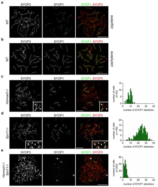
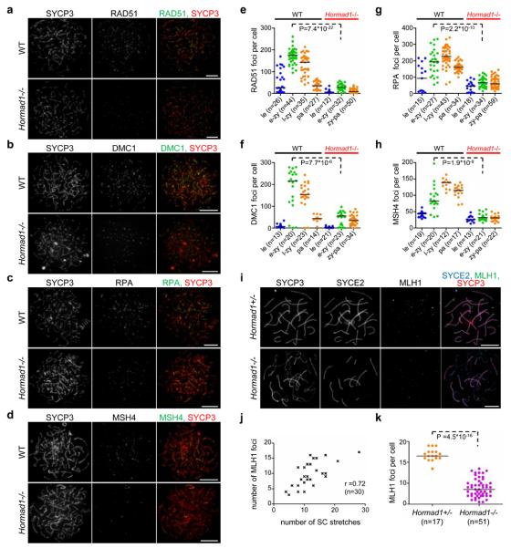
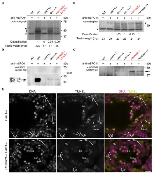
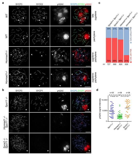
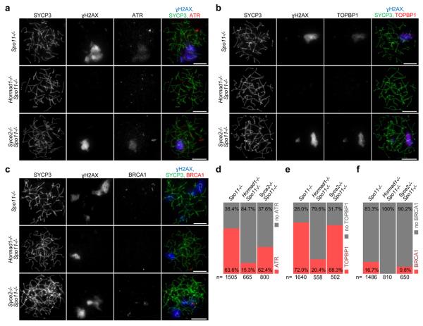
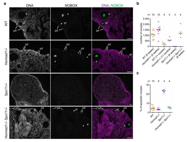
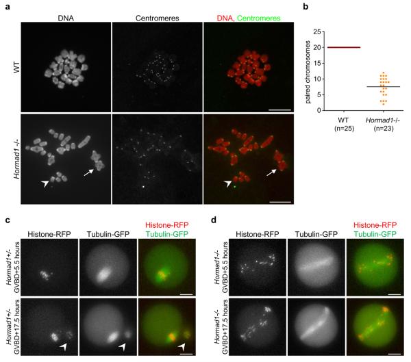
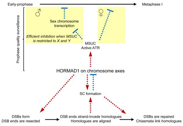
Meiotic crossover formation between homologous chromosomes (homologues) entails DNA double-strand break (DSB) formation, homology search using DSB ends, and synaptonemal-complex formation coupled with DSB repair. Meiotic progression must be prevented until DSB repair and homologue alignment are completed, to avoid the formation of aneuploid gametes. Here we show that mouse HORMAD1 ensures that sufficient numbers of processed DSBs are available for successful homology search. HORMAD1 is needed for normal synaptonemal-complex formation and for the efficient recruitment of ATR checkpoint kinase activity to unsynapsed chromatin. The latter phenomenon was proposed to be important in meiotic prophase checkpoints in both sexes. Consistent with this hypothesis, HORMAD1 is essential for the elimination of synaptonemal-complex-defective oocytes. Synaptonemal-complex formation results in HORMAD1 depletion from chromosome axes. Thus, we propose that the synaptonemal complex and HORMAD1 are key components of a negative feedback loop that coordinates meiotic progression with homologue alignment: HORMAD1 promotes homologue alignment and synaptonemal-complex formation, and synaptonemal complexes downregulate HORMAD1 function, thereby permitting progression past meiotic prophase checkpoints.


Figures

Figure 1
Figure 1. HORMAD1 promotes SC formation independently of DSB-dependent processes
Images of SYCP3 (chromosome axis) and SYCP1 (SC transverse filament), detected by immunofluorescence (IF) on nuclear spreads of WT zygotene (a), WT pachytene (b) and mutant zygotene-pachytene spermatocytes (c-e left) collected from 14-weeks-old mice. The frequency distribution of SC stretches in mutant zygotene-pachytene spermatocytes is shown (c-e right). c, SC formation is never completed on all autosomes in Hormad1−/− cells, and unlike in SC transverse filament mutant meiocytes, where unsynapsed chromosomes align along their length-, unsynapsed chromosomes do not align in Hormad1−/− spermatocytes. Nevertheless, robust stretches of SC frequently form between chromosomes that appear homologous based on their similar axis length. See enlarged view of boxed partially synapsed autosome: unsynapsed axes (arrowheads) are of similar lengths. d, In contrast, SCs connect multiple non-homologous axes, thereby creating a meshwork of interconnected axes in Spo11−/− mutant, in which strand invasion and homology search is not possible due to lack of DSBs. See enlarged view of boxed chromosome axes: arrowheads mark unsynapsed, arrows mark synapsed axes. e. Both the number and the length of SYCP1 stretches are reduced in Hormad1−/− Spo11−/− spermatocytes relative to the single mutants. The majority (61% in n=144 cells) of the remaining SYCP1 stretches is unambiguously linked to a single chromosome axis (arrowheads) indicating that SYCP1 stretches do not necessarily mark inter-chromosomal SCs. Bars, 10 μm.
Figure 2
Figure 2. Numbers of early, intermediate and late recombination protein foci are reduced in the absence of HORMAD1 in prophase meiocytes
a-d, SYCP3 and either RAD51 (a) DMC1 (b), RPA (c) or MSH4 (d) are detected on nuclear spreads of typical early-mid-zygotene WT and Hormad1−/− spermatocytes from 16-days-old mice. Bars, 10 μm. e-h, Foci numbers of early (RAD51-e, DMC1-f) and intermediate (RPA-g, MSH4-h) recombination proteins during leptotene (le) and early-mid-zygotene (e-zy) in WT and Hormad1−/−; late-zygotene (l-zy) and pachytene (pa) in WT and zygotene-pachytene (zy-pa) in Hormad1−/− spermatocytes. Median foci numbers are marked. During the comparable early-mid-zygotene stage, a three- to six-fold reduction (highly significant by Mann–Whitney test) is observed in recombination protein foci numbers in the mutant relative to WT. i-k, Focus numbers of the CO marker MLH1 are reduced in the absence of HORMAD1 in oocytes. i, SYCP3 (chromosome axis), SYCE2 (SC central element) and MLH1 were detected by IF in nuclear spreads of Hormad1+/− and Hormad1−/− oocytes from 19.5 dpc foetuses (a stage when most oocytes are in the late-pachytene or diplotene stage in WT). Fewer chromosome axis-associated MLH1 foci are detected in Hormad1−/− oocytes than in Hormad1+/− oocytes. Note that the majority of MLH1 foci (78%, n=51 cells) are observed on synapsed axes in the mutant. Bars, 10 μm. j, Scatter plot shows positive correlation (Spearman's r=0.72, n=30) between MLH1 foci numbers and the number of SC stretches (immunostaining for SYCE1 or SYCE2) in Hormad1−/− oocytes, indicating that DSBs might be repaired as COs preferentially in chromosome regions that align and synapse, or that synapsis occurs preferentially where COs are successfully designated. k, Chromosome axis-associated MLH1 focus numbers are reduced approximately three-fold in Hormad1−/− oocytes from 18.5-19.5 dpc foetuses relative to Hormad1+/− oocytes. Median focus numbers are marked by horizontal lines.
Figure 3
Figure 3. Amounts of SPO11-oligonucleotide complexes in testes are reduced in the absence of HORMAD1
a, c, Measurement of SPO11-oligonucleotide complexes in testes of adult 14-weeks-old (a) and juvenile 14 dpp (c) mice. SPO11-oligonucleotide complexes were immunoprecipitated with or without anti-SPO11 antibodies, and covalently-linked oligonucleotides were radioactively labelled. Each sample represents one testis-equivalent SPO11-oligonucleotide complexes. Bars mark SPO11-specific signals and asterisks indicate non-specific labelling of a contaminant in the terminal deoxytransferase preparations. Arrowhead marks an artifactual radioactive signal attributable to presence of immunoglobulin heavy chain (Ig-hc in b). Quantified radioactive signals in the mutants have been background-corrected and normalised: in a signals are normalised to the adult WT control; in c Dmc1−/− and Hormad1−/− signals are normalised to their litter mate Dmc1+/+ and Hormad1+/+ controls, respectively (see Methods). Blots of immunoprecipitates from a and c were probed with anti-SPO11 antibodies in b and d, respectively. b, In WT adults, two alternative forms of SPO11 (α and β) are present. Total SPO11 amounts are similar in Dmc1−/− and Hormad1−/− Dmc1−/− mutants, and are lower in the mutants than in WT. Only SPO11β, the form that appears early in meiosis, is detected in the mutants. Arrowhead marks the immunoglobulin heavy chain (bleached out signal). Mid-pachytene spermatogenic block in the mutants (e) is the likely cause of reduced SPO11 amounts in Dmc1−/− and Hormad1−/− Dmc1−/− testes, and of reduced SPO11-oligonucleotide amounts in Dmc1−/− testes, , . d, In juveniles, total SPO11 protein levels are low and only the long β form of SPO11 (arrow) is detectable. For full scan gel-images of a-d see Supplementary Information, Fig, S10. e, DNA was detected by DAPI, and apoptosis was detected by IF-TUNEL assay on cryosections of testes of 15-weeks-old mice. Dmc1−/− and Hormad1−/− Dmc1−/− spermatocytes undergo apoptosis in stage IV tubules as identified by the concomitant presence of intermediate spermatogonia (In), late-prometaphase intermediate spermatogonia (La-In), mitotic intermediate spermatogonia (m) and spermatogonia B (SgB). Both non-apoptotic (sc) and apoptotic (asc) spermatocytes are present in the stage IV tubules shown. Spermatocytes are fully eliminated upon progression to stage V (data not shown). Bars, 20 μm.
Figure 4
Figure 4. HORMAD1 is required for sex body and pseudo-sex body formation in the Spo11+/+ and Spo11−/− backgrounds, respectively
a, SYCP3 (chromosome axis), SYCE2 (SC central element) and γH2AX were detected in nuclear spreads of WT late-zygotene and pachytene, and Hormad1−/− zygotene-pachytene spermatocytes collected from 16-days-old mice. Matched exposure images of γH2AX are shown. In WT late-zygotene spermatocytes, γH2AX chromatin domains associate with unsynapsed chromosome axes. In WT pachytene cells, unsynapsed regions of X and Y sex chromosome axes (marked by x and y) are surrounded by one large γH2AX-rich chromatin domain, the sex body. Anti-γH2AX staining is patchy in the majority (59%) of Hormad1−/− spermatocytes, with no clear correlation between lack of synapsis and γH2AX localisation (third row). A few large γH2AX-rich chromatin domains form in a minority of Hormad1−/− spermatocytes (41%, n=512), but only a subset of unsynapsed axes overlap with γH2AX-rich chromatin, and synapsed axes overlapping with γH2AX-rich chromatin domains are also observed regularly (bottom row). Arrowheads mark two unsynapsed axes in WT late-zygotene and in each mutant cell. Bars, 10 μm. b, Matched exposure images of SYCP3 (chromosome axis), SYCP1 (SC transverse filament) and γH2AX in nuclear spreads of Spo11−/− , Hormad1−/− Spo11−/− and Syce2−/− Spo11−/− spermatocytes of adult (9-weeks-old) mice. Large γH2AX-rich chromatin domains-pseudo-sex bodies (marked by arrowheads) frequently form in Spo11−/− and Syce2−/− Spo11−/− spermatocytes. Bars, 10 μm. c, Quantification of pseudo-sex body formation in spermatocytes with full-length chromosome axes (collected from 24-days-old mice). The percentage of cells with no pseudo-sex body (cells without PSB) or with one to three clear pseudo-sex bodies (cells with PSB) is shown. d, Quantification of γH2AX signal in spermatocytes with full-length chromosome axes (collected from 24-days-old mice) shows a significant reduction in total nuclear γH2AX amounts in Hormad1−/− Spo11−/− relative to Spo11−/− and Syce2−/− Spo11−/− (Mann–Whitney test).
Figure 5
Figure 5. I HORMAD1 is required for efficient accumulation of ATR, TOPBP1 and BRCA1 on chromatin in the absence of programmed DSBs
Matched exposure images of SYCP3 (chromosome axis), γH2AX and either ATR (a), TOPBP1 (b) or BRCA1 (c) in nuclear spreads of spermatocytes collected from 24-days-old mice. Spo11−/− and Syce2−/− Spo11−/− spermatocytes are shown with cloud-like ATR (a) or TOPBP1 (b) accumulation or with BRCA1 localised to axes (c) in γH2AX-marked pseudo-sex bodies. In a and b, Hormad1−/− Spo11−/− cells are shown without a pseudo-sex body and without ATR or TOPBP1 accumulation on chromatin, respectively. In c, Hormad1−/− Spo11−/− cell is shown with a pseudo-sex body, within which no BRCA1 association with the axis was detected. Note that only a small minority of Hormad1−/− Spo11−/− spermatocytes shows pseudo-sex body-like accumulation of γH2AX (Fig. 4c). Bars, 10 μm. d-f, Frequency of cloud-like ATR (d) and TOPBP1 (e) accumulation and frequency of BRCA1 association with axes (f) in spermatocytes with fully formed chromosome axes. ATR- and TOPBP1-rich chromatin domains are frequently observed in Spo11−/− and Syce2−/− Spo11−/− spermatocytes, and BRCA1 association with axes is also observed in these mutants. ATR and TOPBP1 are virtually absent from chromatin in the large majority of Hormad1−/− Spo11−/− cells, and BRCA1 localisation to axes was never observed in Hormad1−/− Spo11−/− spermatocytes.
Figure 6
Figure 6. I Lack of HORMAD1 allows survival of oocytes in the SC-defective Spo11−/− mutant
a, NOBOX (postnatal oocyte marker) was detected by IF on cryosections of ovaries from 6-weeks-old mice. DNA was detected by DAPI. Oocytes in primordial (pd), primary (pr) and secondary (s) follicular stages are shown in WT, Hormad1−/− and Hormad1−/− Spo11−/− ovaries. In the SC-defective Spo11−/− mutant, oocyte numbers are strongly reduced. A lower magnification section of a Spo11−/− ovary is shown to better illustrate the absence of oocytes. Bars, 50 μm. b, Sum of oocyte numbers on every tenth section of sectioned-through ovary pairs at the indicated ages. Each data point represents a mouse. c, Fraction of apoptotic oocytes in the ovaries of 1-day-old (1 dpp) mice of indicated genotypes. Horizontal lines show medians in b and c.
Figure 7
Figure 7. Reduced numbers of chiasmata form in Hormad1−/− oocytes
a, Centromeres were detected by IF and DNA was detected by propidium iodide on nuclear spreads of in vitro-matured metaphase stage oocytes. In WT cells, 20 pairs of chromosomes are connected by chiasmata. In the Hormad1−/− mutant, a large fraction of chromosomes does not have chiasmata (one such chromosome is marked by arrowhead). Arrow marks a pair of chromosomes connected via a chiasma in the mutant oocyte. Note that bivalents are symmetrical and chiasmata form between chromosomes of identical length in the mutant, indicating that CO formation took place between homologous chromosomes. Chromosomes scatter over a larger area during nuclear spreading in the mutant, therefore it was not possible to include all 40 chromosomes of a meiosis I oocyte in the image. Bars, 10 μm. b, The average numbers of paired chromosomes connected by chiasmata (marked by line) are reduced nearly three-fold in in vitro-matured Hormad1−/− oocytes relative to WT oocytes. c and d, mRNAs encoding β-tubulin-GFP and histone H2B-RFP were injected into WT (c) and Hormad1−/− (d) oocytes during the germinal vesicle stage (prophase), and the oocytes were matured in vitro. The fluorescent proteins in the oocytes were imaged 5.5 hours after germinal vesicle break down (GVBD), at a time when WT oocytes are in the first meiotic metaphase, and 17.5 hours after GVBD, at a time when WT oocytes are arrested in the second meiotic metaphase. A polar body (marked by arrowhead in c), which was extruded 7 hours after GVBD (Supplementary Information, Movie 1) is observed next to the metaphase II stage WT oocyte at the 17.5 hour time point. In the Hormad1−/− oocyte (d), the meiotic spindle is abnormally long and chromosomes fail to align at both time points. Meiotic anaphase did not take place in the displayed Hormad1−/− oocyte (Supplementary Information, Movie 2), and no polar body can be observed at either time points (d). Bars, 20 μm.
Figure 8
Figure 8. Model for meiotic progression: negative feedback-loop of HORMAD1 and SC coordinates homology search and meiotic progression
Processes, activation-promotion and inhibition are marked by continuous black arrows, red dashed arrows and blue flat-ended dashed arrows, respectively. HORMAD1 associates with forming chromosome axes at the beginning of meiosis where it promotes DSB formation and/or processing of DSBs. It thereby ensures that adequate numbers of single-stranded DSBs are available for homology search. As part of the homology search process DSB ends strand-invade into homologues. Multiple strand invasion events along the length of chromosomes lead to full alignment of pairs of homologues, which is a prerequisite for the completion of SC formation. HORMAD1 also promotes SC formation through a mechanism that is independent of homology search. SC formation leads to depletion of HORMAD1 from axes and down-regulation of HORMAD1 function. Hence, in spermatocytes, full autosomal SC formation leads to a restriction of HORMAD1 and ATR activity to sex chromosomes, thereby promoting efficient silencing of sex chromosomes, which is a prerequisite for progression beyond mid-pachytene, , . In oocytes, completion of SC formation on all chromosomes leads to complete inactivation of HORMAD1, which in turn leads to down-regulation of ATR and MSUC. Since sustained ATR activity and/or sustained MSUC is believed to block progression beyond meiotic prophase, full SC formation and HORMAD1 inactivation link successful homologue alignment with progression beyond meiotic prophase. Note that successful DSB repair is probably also required for full down-regulation of ATR activity and for meiotic progression in oocytes (for the sake of simplicity, this branch of the prophase checkpoint is not displayed in the model).

References

    1. Baudat F, de Massy B. Regulating double-stranded DNA break repair towards crossover or non-crossover during mammalian meiosis. Chromosome Res. 2007;15:565–577. - PubMed
    1. Hunter N. Meiotic recombination in Molecular genetics of recombination. Springer-Verlag; Berlin Heidelberg: 2007.
    1. Keeney S, Giroux CN, Kleckner N. Meiosis-specific DNA double-strand breaks are catalyzed by Spo11, a member of a widely conserved protein family. Cell. 1997;88:375–384. - PubMed
    1. Baudat F, Manova K, Yuen JP, Jasin M, Keeney S. Chromosome synapsis defects and sexually dimorphic meiotic progression in mice lacking Spo11. Mol Cell. 2000;6:989–998. - PubMed
    1. Romanienko PJ, Camerini-Otero RD. The mouse Spo11 gene is required for meiotic chromosome synapsis. Mol Cell. 2000;6:975–987. - PubMed

Publication types