

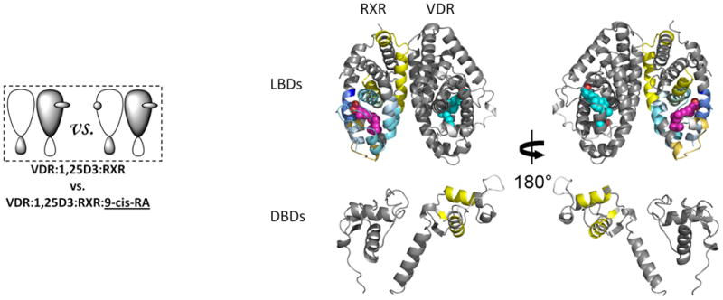
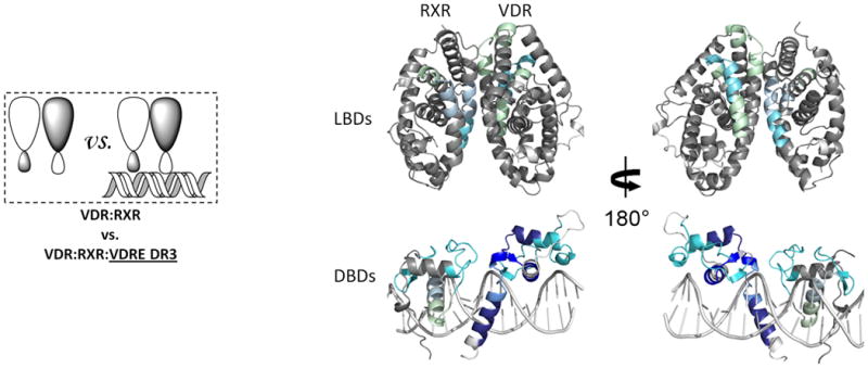
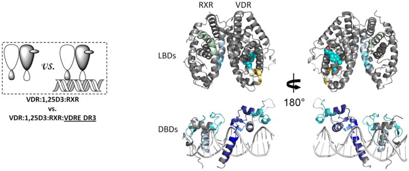
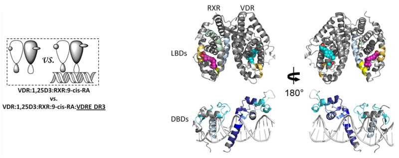
DNA binding alters coactivator interaction surfaces of the intact VDR-RXR complex
- PMID: 21478866
- PMCID: PMC3087838
- DOI: 10.1038/nsmb.2046
DNA binding alters coactivator interaction surfaces of the intact VDR-RXR complex
Abstract
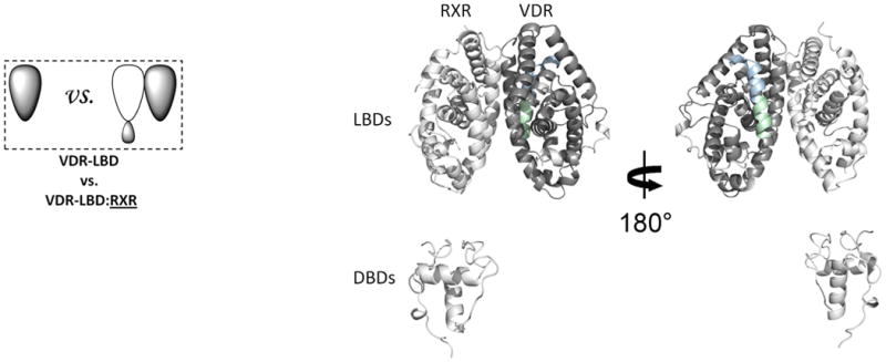
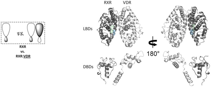
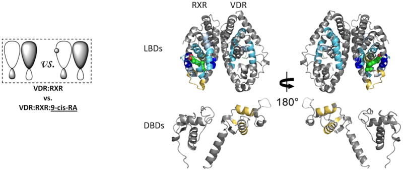
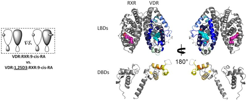
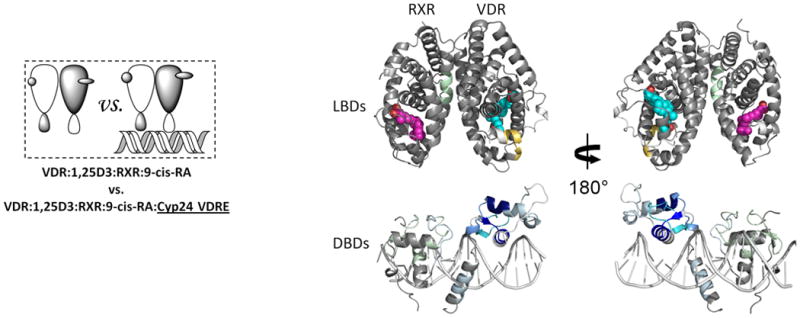
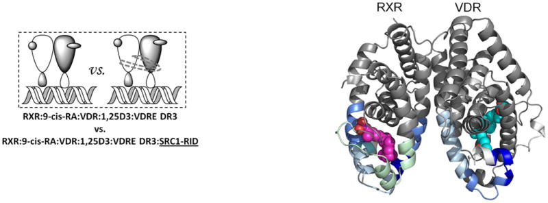
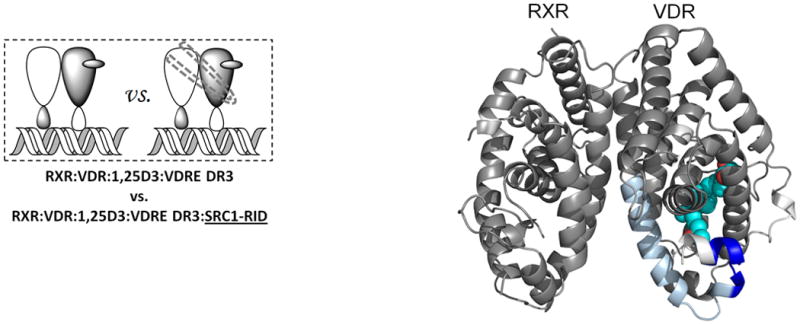
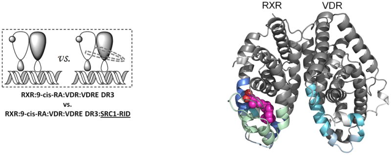
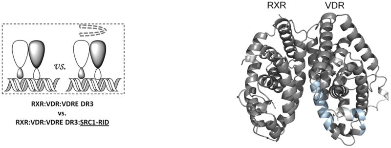
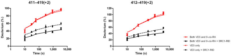
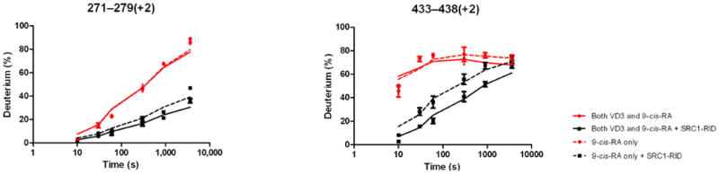
The vitamin D receptor (VDR) functions as an obligate heterodimer in complex with the retinoid X receptor (RXR). These nuclear receptors are multidomain proteins, and it is unclear how various domains interact with one another within the nuclear receptor heterodimer. Here, we show that binding of intact heterodimer to DNA alters the receptor dynamics in regions remote from the DNA-binding domains (DBDs), including the coactivator binding surfaces of both co-receptors, and that the sequence of the DNA response element can determine these dynamics. Furthermore, agonist binding to the heterodimer results in changes in the stability of the VDR DBD, indicating that the ligand itself may play a role in DNA recognition. These data suggest a mechanism by which nuclear receptors show promoter specificity and have differential effects on various target genes, providing insight into the function of selective nuclear receptor modulators.
Figures

References
-
- Gennari L, Merlotti D, De Paola V, Martini G, Nuti R. Update on the pharmacogenetics of the vitamin D receptor and osteoporosis. Pharmacogenomics. 2009;10:417–433. - PubMed
-
- Kostner K, et al. The relevance of vitamin D receptor (VDR) gene polymorphisms for cancer: a review of the literature. Anticancer Res. 2009;29:3511–3536. - PubMed
Publication types
MeSH terms
Substances
Grants and funding
LinkOut - more resources
Full Text Sources
Other Literature Sources
Miscellaneous
