Review
. 2011 May;9(5):369-81.
doi: 10.1038/nrmicro2548. Epub 2011 Apr 11.

Fusing structure and function: a structural view of the herpesvirus entry machinery

Affiliations
Review

Fusing structure and function: a structural view of the herpesvirus entry machinery

Sarah A Connolly et al. Nat Rev Microbiol. 2011 May.

Abstract

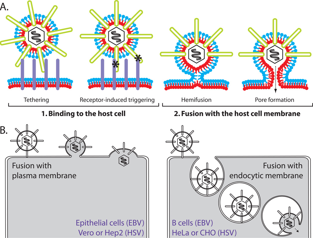
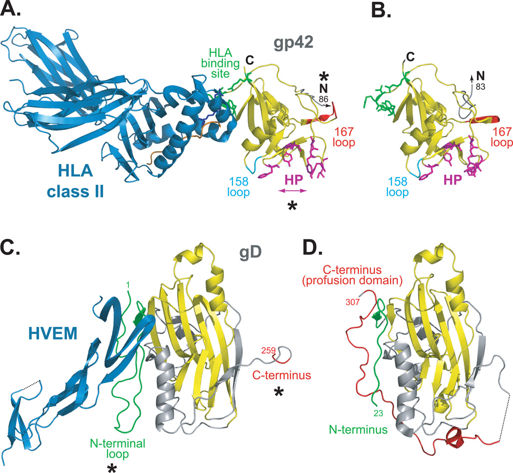
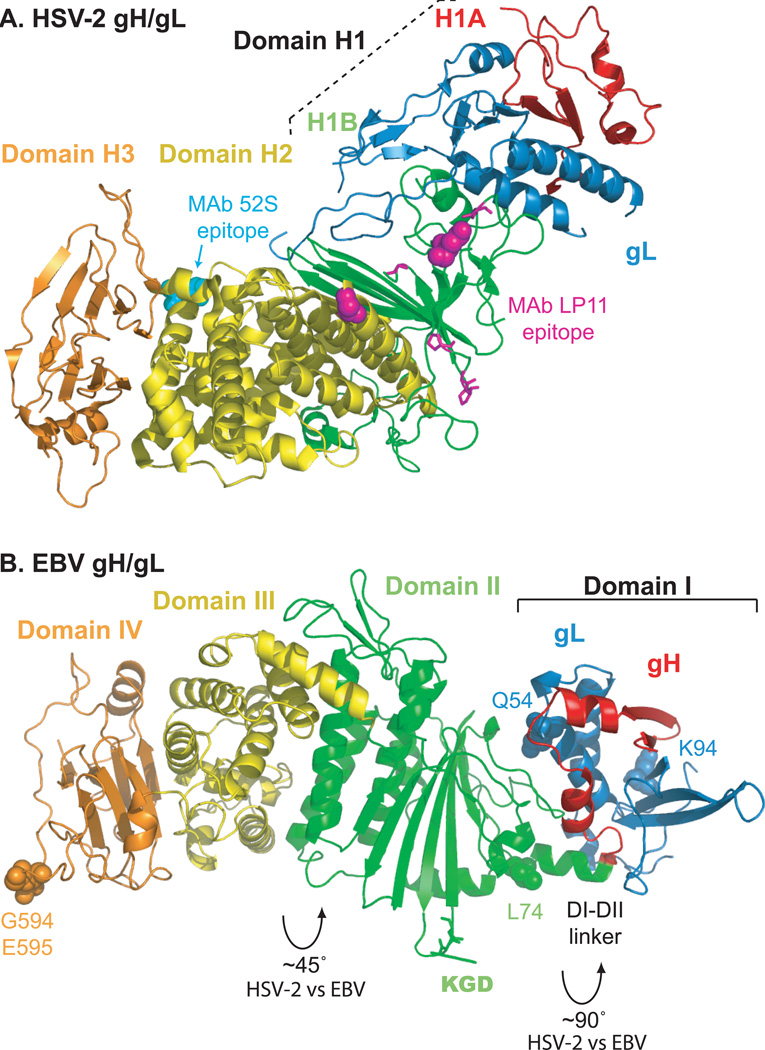
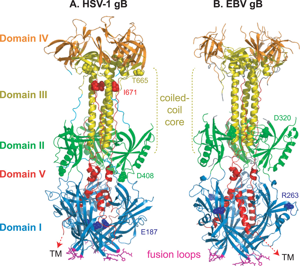
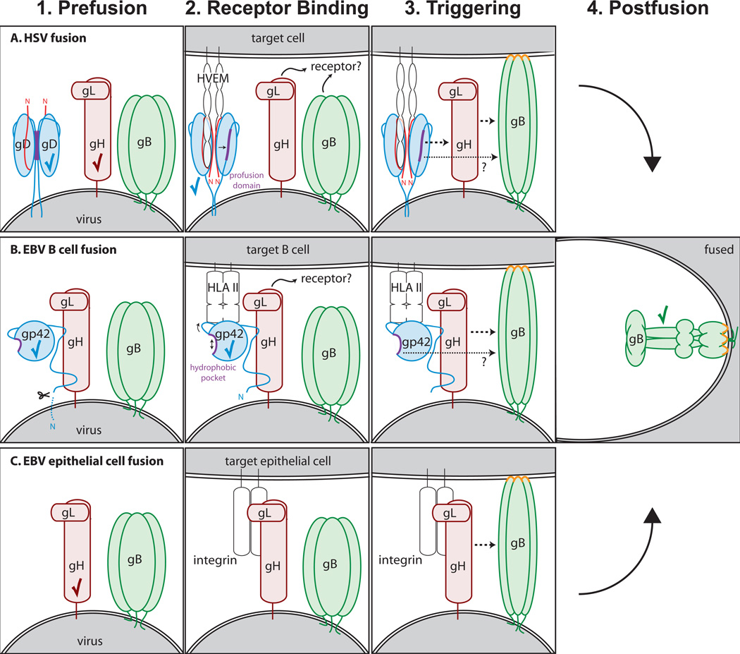
Herpesviruses are double-stranded DNA, enveloped viruses that infect host cells through fusion with either the host cell plasma membrane or endocytic vesicle membranes. Efficient infection of host cells by herpesviruses is remarkably more complex than infection by other viruses, as it requires the concerted effort of multiple glycoproteins and involves multiple host receptors. The structures of the major viral glycoproteins and a number of host receptors involved in the entry of the prototypical herpesviruses, the herpes simplex viruses (HSVs) and Epstein-Barr virus (EBV), are now known. These structural studies have accelerated our understanding of HSV and EBV binding and fusion by revealing the conformational changes that occur on virus-receptor binding, depicting potential sites of functional protein and lipid interactions, and identifying the probable viral fusogen.


Figures

Figure 1
Figure 1. Herpesvirus entry
(A) Routes of entry. Depending upon cell type, both HSV and EBV can enter cells by fusion at the plasma membrane or fusion with an endocytic membrane after endocytosis. Examples of common cells for which each entry route is used are noted. (B) Two steps of virus entry. 1. Virus binds to cellular receptors (purple) via envelope glycoproteins (green). Some receptor binding events serve simply to tether the virus to the cell. Other specific receptor binding events trigger conformational changes in the entry glycoproteins that mediate membrane fusion (asterisks). 2. Fusion of the viral and cellular membranes progresses through a hemifusion intermediate, in which the outer membrane leaflets (blue) mix. This is followed by full fusion, where the inner membrane leaflets (red) mix and a fusion pore is formed. This figure is not drawn to scale.
Figure 2
Figure 2. EBV and HSV receptor binding proteins, in bound and unbound states
(A) EBV gp42 bound to class II HLA-DR1 (PDB ID 1KG0). (B) Unbound gp42 (PDB ID 3FD4). gp42 binds HLA class II (blue) at a non-canonical site within the gp42 CTLD (yellow, residues 94–221). HLA class II residues E46 and R72 (blue sticks) make extensive contacts with gp42 residues 104–107 and R200 (green sticks). The canonical CTLD interaction site lies on an opposite face of gp42 at a pocket lined with hydrophobic residues (magenta sticks). Asterisks denote conformational differences seen in gp42 in the presence or absence of HLA class II. When bound to HLA, gp42 loops at residue 158 (cyan) and 167 (red) are shifted and the hydrophobic pocket (HP) widens compared to unbound gp42. In the absence of HLA, the gp42 N-terminus projects outward in a path distinct from that observed for HLA-bound gp42. The N-terminus of gp42 is flexible and the residues that bind to gH/gL (gp42 residues 36–81) were not resolved. Peptide loaded in the HLA class II is colored orange. (C) HSV-1 gD bound to HVEM (PDB ID 1JMA). A truncated form of the gD ectodomain (gD285) with high affinity for HVEM was crystallized in complex with HVEM (blue). (D) Unbound gD (PDB ID 2C3A). A cysteine was added to the C-terminus of the gD ectodomain [gD(23–306)307C] which stabilized a gD dimer. A monomer is shown. The core of gD forms an Ig fold (yellow) that is flanked by N-terminal (green) and C-terminal (red) extensions. Asterisks denote conformational differences seen in gD in the presence or absence of receptor. In the presence of HVEM, the N-terminus of gD (green) forms a loop that serves as the receptor binding site and is stabilized by receptor binding. The C-terminal region, residues 260–285, is disordered in the crystal. In the absence of HVEM, the C-terminal region (red), which includes the pro-fusion domain, is anchored near the N-terminal region (green). The C-terminus masks the receptor binding site and the N-terminal loop conformation is not present. The structure of gD285 (PDB ID 1L2G, not shown) in the absence of receptor demonstrates a similar loss of the N-terminal loop and its N-terminus is disordered past residue 14 .
Figure 3
Figure 3. Conserved entry glycoproteins gH/gL
(A) HSV-2 gH/gL (PDB ID 3M1C). The HSV gH/gL heterodimer adopts a boot-like configuration. The N-terminal gH domain H1, comprised of subdomains H1A (red) and H1B (green), clamps gL (blue) and make extensive contacts. The C-terminal gH domains H2 (yellow) and H3 (orange) are shown. Substitution mutations at gH residues 168 or 329 (magenta spheres) or insertion mutations at gH residues 300, 313, 316, or 317 (magenta sticks) prevent binding of the neutralizing MAb LP11. On the opposite face of the complex, substitution mutations at gH 536 or 537 (cyan spheres) prevent binding of the neutralizing MAb 52S. (B) EBV gH/gL (PDB ID 3PHF). EBV gH/gL domain designations differ somewhat from HSV-2. Domain I is comprised of gL (blue) and the N-terminus of gH (red). Domain II (green) contains an eight-stranded β-sheet that forms a ‘picket fence’ separating domain I from a helical bundle within domain II that lies parallel to the fence. Domain III (yellow) is mainly helical and domain IV (orange) forms a β-sandwich. The EBV gH/gL heterodimer is more linear and elongated than the HSV-2 complex because the interdomain packing arrangements of EBV gH/gL differ significantly. Packing angles differ by ~90° between domains I and II and ~45° between domains II and III. EBV gL residues Q54 and K94 (blue spheres) contribute to a functional interaction with EBV gB. A KGD motif (green sticks) is located in a prominent loop of domain II and lies adjacent to the helical linker between domains I and II. A mutation at residue gH-L74 (green spheres) near the linker results in increased fusion promotion. Mutations at gH residues G594 or E595 (orange spheres) differentially affect fusion promotion.
Figure 4
Figure 4. Conserved fusion protein gB
(A) HSV-1 gB (PDB ID 2GUM). (B) EBV gB (PDB ID 3FVC). Domain I (blue) contains hydrophobic fusion loops (magenta) that insert into the target cell membrane. For crystallization, the hydrophobic residues in the EBV gB fusion loops were replaced with corresponding residues from HSV-1 gB. The C-terminal domain V (red) packs against the coiled-coil core formed by domain III (yellow) and proceeds through domain I, headed towards the transmembrane (TM) region. This creates a hairpin-like organization of the structure wherein the fusion loops and TM lie at the same end of the trimer. The location of insertion mutations that reduce fusion without preventing gB expression are noted (spheres colored according to domain).
Figure 5
Figure 5. Models of fusion
(A) HSV fusion with a target cell at the plasma or endosomal membrane. 1. gD is expressed as a dimer with its C-termini occluding its receptor binding site. 2. Upon binding HVEM, the N-terminus of gD (red) forms a loop and displaces the gD C-terminus that contains the profusion domain (purple). Nectin-1 binds to an overlapping site on gD and displaces its C-terminus in a similar manner. The gB trimer also possesses receptor-binding activity and the gH/gL heterodimer may bind to a cellular receptor as well. 3. Different sites within the gD profusion domain can interact with gH/gL and gB. In addition, gH/gL and gB may interact with one another in response to gD binding receptor. These interactions trigger gB to insert its fusion loops (orange) into the target membrane. 4. gB then refolds to its postfusion conformation to mediate fusion of the viral and cellular membranes. (B) EBV fusion with B cell endosomal membrane. 1. gp42, a type II glycoprotein, undergoes N-terminal cleavage and is retained on the virion by binding gH/gL via the gp42 N-terminus. 2. When HLA class II binds to gp42, a hydrophobic pocket (purple) on gp42 widens. gH/gL may also bind a receptor. 3. Conformational changes within gp42 and/or gH/gL trigger gB fusion loop (orange) insertion. The hydrophobic pocket in gp42 may serve as an interaction site to promote fusion. 4. gB refolds to a postfusion conformation, thereby mediating membrane fusion. (C) EBV fusion with epithelial cell plasma membrane. 1. gp42 functions as a tropism switch and inhibits entry into epithelial cells. Virus produced in B cells is deficient in gp42 and thus able to infect epithelial cells. 2. gH/gL binds to receptor such as integrin. 3. After receptor binding, gH/gL interacts with gB and triggers fusion loop (orange) insertion. 4. gB refolds to a postfusion conformation, thereby driving membrane fusion. Checkmarks denote structures that have been solved. Note that gp42 and gH/gL were not solved in complex. Structures for the other protein complexes and conformations, such as receptor-bound gH/gL and pre-fusion gB, remain to be determined.

References

    1. Pellet PE, Roizman B. In: Fields' Virology. 5th Edition. Knipe DM, Howley PM, editors. New York, N.Y: Lippincott-Williams and Wilkins; 2007. pp. 2479–2499.
    1. Milne RS, Nicola AV, Whitbeck JC, Eisenberg RJ, Cohen GH. Glycoprotein D receptor-dependent, low-pH-independent endocytic entry of herpes simplex virus type 1. J Virol. 2005;79:6655–6663. - PMC - PubMed
    1. Nicola AV, McEvoy AM, Straus SE. Roles for endocytosis and low pH in herpes simplex virus entry into HeLa and Chinese hamster ovary cells. J Virol. 2003;77:5324–5332. - PMC - PubMed
    1. Dollery SJ, Delboy MG, Nicola AV. Low pH-induced conformational change in herpes simplex virus glycoprotein B. J Virol. 2010;84:3759–3766. - PMC - PubMed
    1. Stampfer SD, Lou H, Cohen GC, Eisenberg RJ, Heldwein EE. Structural basis of local, pH-dependent conformational changes in glycoprotein B from herpes simplex virus 1. J Virol. 2010 - PMC - PubMed

Publication types