. 2010 Apr 11;3(3):127-39.
doi: 10.1007/s12154-010-0038-2.

GroEL-induced topological dislocation of a substrate protein β-sheet core: a solution EPR spin-spin distance study

GroEL-induced topological dislocation of a substrate protein β-sheet core: a solution EPR spin-spin distance study

Rikard Owenius et al. J Chem Biol. .

Abstract

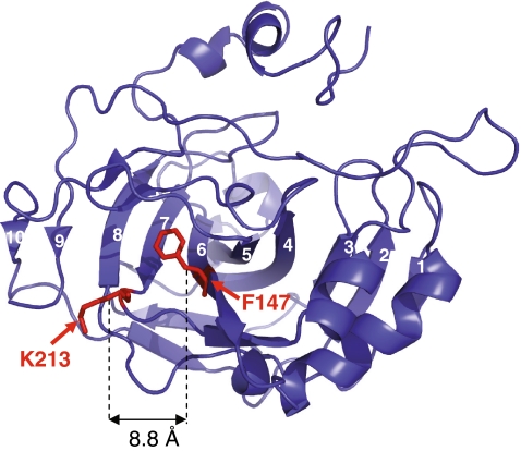
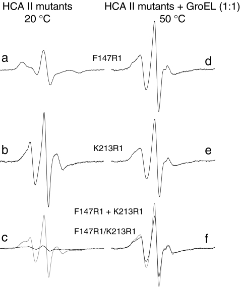
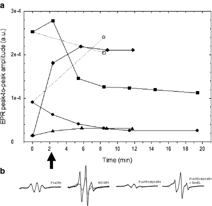
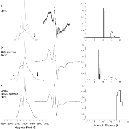
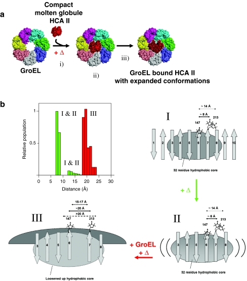
The Hsp60-type chaperonin GroEL assists in the folding of the enzyme human carbonic anhydrase II (HCA II) and protects it from aggregation. This study was aimed to monitor conformational rearrangement of the substrate protein during the initial GroEL capture (in the absence of ATP) of the thermally unfolded HCA II molten-globule. Single- and double-cysteine mutants were specifically spin-labeled at a topological breakpoint in the β-sheet rich core of HCA II, where the dominating antiparallel β-sheet is broken and β-strands 6 and 7 are parallel. Electron paramagnetic resonance (EPR) was used to monitor the GroEL-induced structural changes in this region of HCA II during thermal denaturation. Both qualitative analysis of the EPR spectra and refined inter-residue distance calculations based on magnetic dipolar interaction show that the spin-labeled positions F147C and K213C are in proximity in the native state of HCA II at 20 °C (as close as ∼8 Å), and that this local structure is virtually intact in the thermally induced molten-globule state that binds to GroEL. In the absence of GroEL, the molten globule of HCA II irreversibly aggregates. In contrast, a substantial increase in spin-spin distance (up to >20 Å) was observed within minutes, upon interaction with GroEL (at 50 and 60 °C), which demonstrates a GroEL-induced conformational change in HCA II. The GroEL binding-induced disentanglement of the substrate protein core at the topological break-point is likely a key event for rearrangement of this potent aggregation initiation site, and hence, this conformational change averts HCA II misfolding.

Keywords: Carbonic anhydrase; Misfolding; Molecular chaperone; Molten globule; Protein aggregation; Unfoldase.


Figures

Fig. 1
Fig. 1
The HCA II structure in cartoon representation with the sites of mutation, residues F147 and K213, indicated in red. The β-carbon–β-carbon distance between these sites is 8.8 Å. The ten central β-strands of HCA II are numbered. β-strands 6 and 7 are parallel and the remainder of the central β-sheet is in an antiparallel arrangement. The figure was generated using PyMol (DeLano Scientific, LLC) with coordinates provided from RCSB Protein Data Bank (accession code 2CBA [39])
Fig. 2
Fig. 2
EPR spectra of spin labeled HCA II mutants in 0.1 M Tris–H2SO4, pH 7.5 at 20 °C: a F147R1, b K213R1, and c F147R1/K213R1 (black trace) and F147R1 + K213R1 (summed spectra) (gray trace). EPR spectra of spin labeled HCA II mutants bound to GroEL at equimolar concentration in 0.1 M Tris–H2SO4, pH 7.5 at 50 °C: d F147R1, e K213R1, and f F147R1/K213R1 (black trace) and F147R1 + K213R1 (summed spectra) (gray trace). The same amplitude scale was used in all spectra, but spectra in c and f had first been divided by 2 to have all spectra representing one-spin systems. The scan width was 120 G
Fig. 3
Fig. 3
a EPR peak-to-peak amplitudes (Aptp) of the mI = 0 hyperfine line as a function of time after the 20 °C sample has been inserted into the resonator, preset to 50 °C: filled circles F147R1, filled square K213R1, filled triangle F147R1/K213R1, and filled diamond F147R1/K213R1 and GroEL, all in 0.1 M Tris–H2SO4, pH 7.5. All Aptp values were normalized to the same parameter set to allow comparison (see “Materials and methods” section). Aptp values at 0 min were taken from spectra measured at 20 °C (Fig. 2). Time values of data obtained at t > 0 correspond to the time when the center of the mI = 0 line was swept. For comparison, Aptp values from single mutants equilibrated at 50 °C in the presence of GroEL were included: open circles GroEL-F147R1 and open squares GroEL-K213R1. Lines were displayed for guidance. b The first one-scan EPR spectra taken after samples were introduced to 50 °C. The scan was started after 1 min and corresponding Aptp values are marked with an arrow in a. For K213R1, the last spectrum taken (after 18 min) is shown for comparison (gray trace). The same y-axis scale was used for all spectra
Fig. 4
Fig. 4
Left part, absorption EPR spectra of F147R1/K213R1 (black trace) and F147R1 + K213R1 (gray trace). All spectra are plotted with reference to a 300 G field scan. The vertical arrows denote regions of spectral broadening due to dipolar interaction. Middle part, first-derivative EPR spectra of F147R1/K213R1 together with simulated spectra from the distance analysis (dashed line). All spectra are plotted with reference to a spectral width of 200 G. Right part, derived interspin distance distributions expressed as normalized distance populations. a At 20 °C with no viscosity modifying agent, b with 40% sucrose at 20 °C, and c with 52.5% sucrose and equimolar amounts of GroEL at 60 °C
Fig. 5
Fig. 5
a Schematic top view structural model showing GroEL binding-induced unfolding of HCA II. i The thermally induced molten globule is captured by multiple valences to the GroEL apical domains. ii Initially, the molten globule has preserved central strand compactness, and iii the GroEL induced expansion of the central core of the bound substrate. b Cartoon representations of the topography of the native (I), molten globule (II), and GroEL bound HCA II (III). Distance distributions between position F147R1 and K213R1 (top left image). The capital roman numerals refer to the conformational states in the cartoon. I In the native state (20 °C) (top right cartoon), the ten stranded β-sheet with antiparallel arrangement between β-strands 1–6 and between β strands 7–9 are indicated, together with the parallel arrangement of β-strands 6 and 7 forming a topological breakpoint. The two calculated spin–spin distance populations of ∼8 Å and ∼14 Å are indicated. II The central core comprising residues from strands 3–8 are preserved in the molten globule state formed at elevated temperature [37] (lower right cartoon), as well as the native state distance between residues F147R1 and K213R1, with distances of ∼8 Å and ∼14 Å from spin–spin EPR measurements. III The GroEL bound state (bottom left cartoon) shows the GroEL-induced separated topological breakpoint indicated by three distance arrows. The short arrow includes the uncertainties of CW EPR distance analysis with deconvolution methods (often limited to 15–17 Å distances [58] the second arrow is taken from our calculated distance distribution ∼20 Å, and the third arrow denotes the possibility that the true distance is >20 Å, where the method is irrefutably blind

Similar articles

Cited by

References

    1. Chiti F, Dobson CM. Protein misfolding, functional amyloid, and human disease. Annu Rev Biochem. 2006;75:333–366. doi: 10.1146/annurev.biochem.75.101304.123901. - DOI - PubMed
    1. Lambert MP, Barlow AK, Chromy BA, Edwards C, Freed R, Liosatos M, Morgan TE, Rozovsky I, Trommer B, Viola KL, Wals P, Zhang C, Finch CE, Krafft GA, Klein WL. Diffusible, nonfibrillar ligands derived from Abeta1-42 are potent central nervous system neurotoxins. Proc Natl Acad Sci USA. 1998;95:6448–6453. doi: 10.1073/pnas.95.11.6448. - DOI - PMC - PubMed
    1. Hammarström P, Persson M, Freskgård PO, Mårtensson LG, Andersson D, Jonsson BH, Carlsson U. Structural mapping of an aggregation nucleation site in a molten globule intermediate. J Biol Chem. 1999;274:32897–32903. doi: 10.1074/jbc.274.46.32897. - DOI - PubMed
    1. Wiseman RL, Powers ET, Buxbaum JN, Kelly JW, Balch WE. An adaptable standard for protein export from the endoplasmic reticulum. Cell. 2007;131(4):809–821. doi: 10.1016/j.cell.2007.10.025. - DOI - PubMed
    1. London J, Skrzynia C, Goldberg ME. Renaturation of Escherichia coli tryptophanase after exposure to 8 M urea. Evidence for the existence of nucleation centers. Eur J Biochem. 1974;47:409–415. doi: 10.1111/j.1432-1033.1974.tb03707.x. - DOI - PubMed