

In Vitro and in Vivo Characteristics of Fluorapatite-Forming Calcium Phosphate Cements
- PMID: 21479080
- PMCID: PMC3072690
- DOI: 10.6028/jres.115.020
In Vitro and in Vivo Characteristics of Fluorapatite-Forming Calcium Phosphate Cements
Abstract
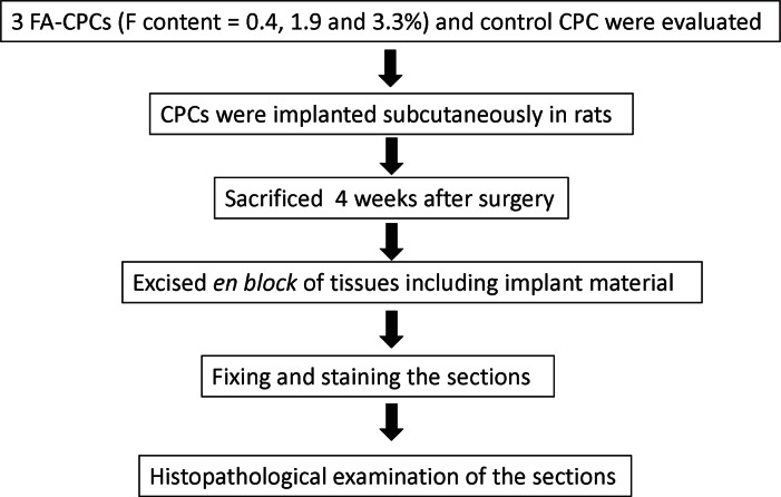
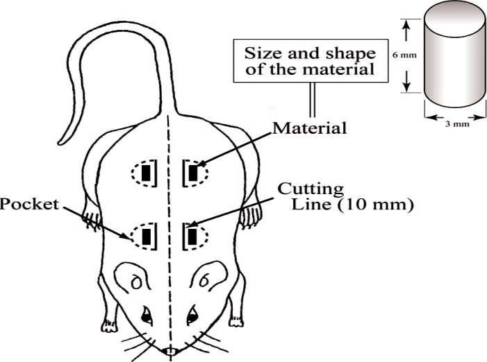
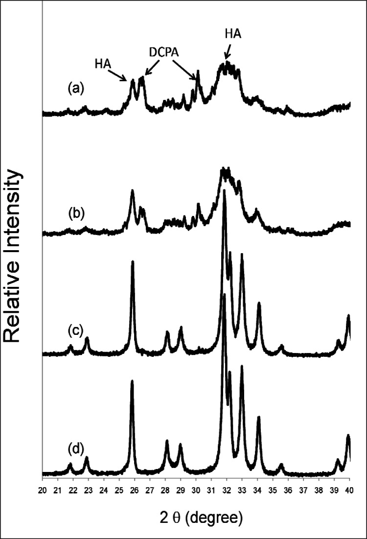
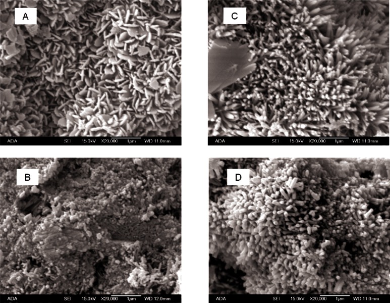
This study reports for the first time in vitro and in vivo properties of fluorapatite (FA)-forming calcium phosphate cements (CPCs). The experimental cements contained from (0 to 3.1) mass % of F, corresponding to presence of FA at levels of approximately (0 to 87) mass %. The crystallinity of the apatitic cement product increased greatly with the FA content. When implanted subcutaneously in rats, the in vivo resorption rate decreased significantly with increasing FA content. The cement with the highest FA content was not resorbed in soft tissue, making it the first known biocompatible and bioinert CPC. These bioinert CPCs might be useful for applications where slow or no resorption of the implant is required to achieve the desired clinical outcome.
Figures


Similar articles
-
Development of a novel fluorapatite-forming calcium phosphate cement with calcium silicate: in vitro and in vivo characteristics.Dent Mater J. 2015;34(2):263-9. doi: 10.4012/dmj.2014-255. Epub 2015 Feb 25. Dent Mater J. 2015. PMID: 25740309
-
Transforming growth factor-beta1 incorporation in a calcium phosphate bone cement: material properties and release characteristics.J Biomed Mater Res. 2002 Feb;59(2):265-72. doi: 10.1002/jbm.1241. J Biomed Mater Res. 2002. PMID: 11745562
-
Effect of a novel fluorapatite-forming calcium phosphate cement with calcium silicate on osteoblasts in comparison with mineral trioxide aggregate.J Oral Sci. 2015 Mar;57(1):25-30. doi: 10.2334/josnusd.57.25. J Oral Sci. 2015. PMID: 25807905
-
Injectable bone cements: What benefits the combination of calcium phosphates and bioactive glasses could bring?Bioact Mater. 2022 Apr 20;19:217-236. doi: 10.1016/j.bioactmat.2022.04.007. eCollection 2023 Jan. Bioact Mater. 2022. PMID: 35510175 Free PMC article. Review.
-
Biological and mechanical performance and degradation characteristics of calcium phosphate cements in large animals and humans.Acta Biomater. 2020 Nov;117:1-20. doi: 10.1016/j.actbio.2020.09.031. Epub 2020 Sep 23. Acta Biomater. 2020. PMID: 32979583 Review.
Cited by
-
Development of nanofluorapatite polymer-based composite for bioactive orthopedic implants and prostheses.Int J Nanomedicine. 2014 Aug 11;9:3875-84. doi: 10.2147/IJN.S65682. eCollection 2014. Int J Nanomedicine. 2014. PMID: 25143735 Free PMC article.
-
Proliferation, odontogenic/osteogenic differentiation, and cytokine production by human stem cells of the apical papilla induced by biomaterials: a comparative study.Clin Cosmet Investig Dent. 2019 Jul 12;11:181-193. doi: 10.2147/CCIDE.S211893. eCollection 2019. Clin Cosmet Investig Dent. 2019. PMID: 31372059 Free PMC article.
-
Nanostructured platforms for the sustained and local delivery of antibiotics in the treatment of osteomyelitis.Crit Rev Ther Drug Carrier Syst. 2015;32(1):1-59. doi: 10.1615/critrevtherdrugcarriersyst.2014010920. Crit Rev Ther Drug Carrier Syst. 2015. PMID: 25746204 Free PMC article. Review.
-
Does translational symmetry matter on the micro scale? Fibroblastic and osteoblastic interactions with the topographically distinct poly(ε-caprolactone)/hydroxyapatite thin films.ACS Appl Mater Interfaces. 2014 Aug 13;6(15):13209-20. doi: 10.1021/am503043t. Epub 2014 Jul 23. ACS Appl Mater Interfaces. 2014. PMID: 25014232 Free PMC article.
-
Self-setting calcium orthophosphate formulations.J Funct Biomater. 2013 Nov 12;4(4):209-311. doi: 10.3390/jfb4040209. J Funct Biomater. 2013. PMID: 24956191 Free PMC article.
References
-
- Brown WE, Chow LC. A new calcium phosphate setting cement. J Dent Res. 1983;62(Spec. Iss.) Abst. No. 207.
-
- Sugawara A, Kusama K, Nishimura S, Nishiyama M, Ohashi M, Moro I, Chow LC, Takagi S. Biocompatibility and osteoconductivity of calcium phosphate cement. J Dent Res. 1990;69(Spec. Iss.) Abst. No. 1628.
-
- Chow LC, Takagi S, Costantino PD, Friedman CD. Self-setting calcium phosphate cements. Mater Res Soc Symp Proc. 1991;179:3–24.
-
- Sugawara A, Nishiyama M, Kusama K, Moro I, Nishimura S, Kudo I, Chow LC, Takagi S. Histopathological reactions of calcium phosphate cement. Dent Mater J. 1992;11:11–16. - PubMed
-
- Sugawara A, Fujikawa K, Kusama K, Nishiyama M, Murai S, Takagi S, Chow LC. Histopathologic reaction of calcium phosphate cement for alveolar ridge augmentation. J Biomed Mater Res. 2002;61:47–52. - PubMed
Grants and funding
LinkOut - more resources
Full Text Sources