. 2011 Mar 30;6(3):e18085.
doi: 10.1371/journal.pone.0018085.

Influence of caloric restriction on constitutive expression of NF-κB in an experimental mouse astrocytoma

Affiliations

Influence of caloric restriction on constitutive expression of NF-κB in an experimental mouse astrocytoma

Tiernan J Mulrooney et al. PLoS One. .

Abstract

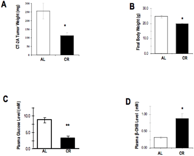
Background: Many of the current standard therapies employed for the management of primary malignant brain cancers are largely viewed as palliative, ultimately because these conventional strategies have been shown, in many instances, to decrease patient quality of life while only offering a modest increase in the length of survival. We propose that caloric restriction (CR) is an alternative metabolic therapy for brain cancer management that will not only improve survival but also reduce the morbidity associated with disease. Although we have shown that CR manages tumor growth and improves survival through multiple molecular and biochemical mechanisms, little information is known about the role that CR plays in modulating inflammation in brain tumor tissue.

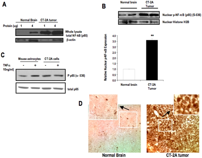
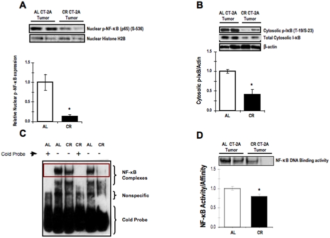
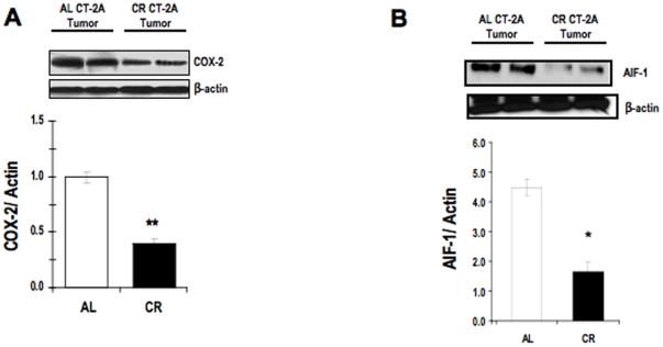
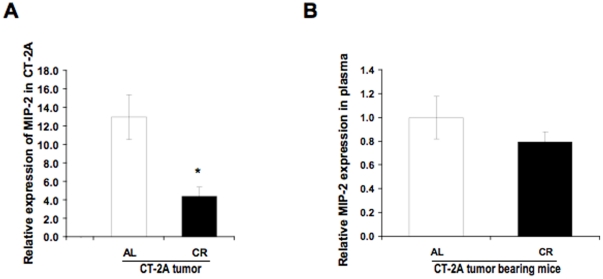
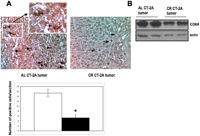
Methodology/principal findings: Phosphorylation and activation of nuclear factor κB (NF-κB) results in the transactivation of many genes including those encoding cycloxygenase-2 (COX-2) and allograft inflammatory factor-1 (AIF-1), both of which are proteins that are primarily expressed by inflammatory and malignant cancer cells. COX-2 has been shown to enhance inflammation and promote tumor cell survival in both in vitro and in vivo studies. In the current report, we demonstrate that the p65 subunit of NF-κB was expressed constitutively in the CT-2A tumor compared with contra-lateral normal brain tissue, and we also show that CR reduces (i) the phosphorylation and degree of transcriptional activation of the NF-κB-dependent genes COX-2 and AIF-1 in tumor tissue, as well as (ii) the expression of proinflammatory markers lying downstream of NF-κB in the CT-2A malignant mouse astrocytoma, [e.g. macrophage inflammatory protein-2 (MIP-2)]. On the whole, our date indicate that the NF-κB inflammatory pathway is constitutively activated in the CT-2A astrocytoma and that CR targets this pathway and inflammation.

Conclusion: CR could be effective in reducing malignant brain tumor growth in part by inhibiting inflammation in the primary brain tumor.


Conflict of interest statement

Competing Interests: The authors have declared that no competing interests exist.

Figures

Figure 1
Figure 1. Expression and Phosphorylation of NF-κB (p65) in CT-2A astrocytoma.
Western blot analysis of (A) Total protein expression of NF-κB (p65) in the whole lysates of tumor tissue and normal brain parenchyma. 1 and 4 µg of protein was loaded for each sample of tumor and brain tissue. (B) Phosphorylated NF-κB (p65) in nuclear extracts of tumor and normal brain tissues. The histogram illustrates the average relative expression of p-NFκB (p65) (S-536) to histone in nuclear extracts of the indicated tissue. Values are expressed as normalized means ± S.E.M of 4–5 independent tissue samples/group for both A and B. The asterisks in indicate that the value is significantly higher in the CT-2A astrocytoma than in contra-lateral normal brain at ** P<0.01 (Student t-test). Two representative samples are shown for each tissue type. (C) Phosphorylation of NF-κB in CT-2A cells and control mouse astrocytes in absence and presence of TNFα (10 ng/ml). (D) NF-κB immunostaining in CT-2A tumor and contra-lateral normal brain tissues. 3 independent mouse brain tumors were analyzed.
Figure 2
Figure 2. Influence of CR on tumor growth, body weight, and plasma biomarkers.
Tumors were implanted i.c. on day 0. All tumor-bearing mice were fed ad libitum (AL) for the first 48 hrs after tumor implantation and were then randomly assigned to one of two diet groups that received rodent chow in either unrestricted (AL; n = 4) or calorically restricted (CR; n = 5) amounts. The two groups were matched for body weight (∼28.0 g) prior to the initiation of CR. The CR group received a feeding regimen of 30% less or 70% of food of AL mice consume daily. All mice were sacrificed 14–15 days after tumor implantation, as described above in the Materials and Methods section. Final weights of the mice were obtained immediately prior to sacrifice (B). The asterisk in indicate that the tumor growth (A), body weight (B), and glucose level (C) is significantly reduced and ketone level (D) is significantly increased in the CR group compared with the AL group at * P<0.05, ** P<0.001 (Student t-test).
Figure 3
Figure 3. Influence of CR on NF-κB expression and activation in CT-2A astrocytoma.
Nuclear expression of phosphorylated NF-κB (p65) (A); cytosolic expression of phosphorylated IκB and total lκB (B) as assessed by western blot analysis, DNA promoter binding activity of activated NF-κB in the CT-2A astrocytoma (C-D) as assessed by EMSA. The histograms illustrate the average relative expression of phosphorylated to total protein normalized to the indicated loading control in either nuclear or cytoplasmic extracts of the indicated tissue (A-B). Values are expressed as normalized means ± S.E.M of 4–5 independent tissue samples/group. The asterisks in indicate that the value is significantly different in the CT-2A astrocytoma under AL and CR condition at * P<0.05 (Student t-test). Two representative samples are shown for each tissue type. (C) Evaluation of the extent of DNA proinflammatory gene promoter binging activity by activated NF-κB in nuclear extracts of CT-2A under AL and CR condition. (D) Coned-down view of the DNA promoter biding activity of activated NF-κB in nuclear extracts of the NF-κB in the CT-2A astrocytoma under AL and CR condition. The histogram illustrates the average relative expression of activated NF-κB in the indicated tissue. Values are expressed as normalized means ± S.E.M of 4–5 independent tissue samples/group. The asterisks indicate that the value is significantly different in the CT-2A astrocytoma under AL and CR condition at * P<0.05 (Student t-test). Two representative samples are shown for each tissue type.
Figure 4
Figure 4. Influence of CR on inflammatory protein expressions in CT-2A astrocytoma.
Cyclooxygenase-2 (COX-2) (A) and Allograft Inflammatory Factor 1 (AIF-1) (B) in cytosolic extracts of the CT-2A astrocytoma. COX-2 and AIF-1 have both been reported to be downstream proinflammatory gene-product effectors of the activated NF-κB. The histograms illustrate the average relative expression of the indicated protein normalized to β-actin in CT-2A tumors (A-B). Values are expressed as normalized means ± S.E.M of 4–5 independent tissue samples/group. The asterisks indicate that the value is significantly different in the CR tumor than in the AL tumor at * P<0.05, † P<0.001 (Student t-test). Two representative samples are shown for each tissue type.
Figure 5
Figure 5. Influence of CR on the Macrophage Inflammatory Protein-2 (MIP-2) concentration.
MIP-2 concentration in brain tumor lysates and in the plasma of mice bearing orthotopically implanted CT-2A tumors as assessed by ELISA. The histograms illustrate the average relative expression of the indicated protein in CT-2A from AL- and CR-fed mice (A). Influence of CR on expression of the indicated protein in the plasma of CT-2A-bearing mice fed AL or CR (B). All values are expressed as means ± S.E.M of 4-5 independent tissues or plasma samples/group. ** P<0.01 indicates that the average MIP-2 level in tumors from the AL-fed group is significantly different from that in tumors from the CR-fed group (Student t-test).
Figure 6
Figure 6. Influence of CR on CD68 expression in CT-2A tumor.
Immunohistochemistry staining shows brown CD68 positive (black arrow) cells at x400 (A). Each section was representative of the entire tumor. All images are produced from digital photograph. The histograms illustrate the average number of brown cells in the entire tumor section at 400× from 3 independent mouse tumors in each group. *P<0.05 indicates that the average CD68 positive cells in tumors from the CR-fed group is significantly less from that in tumors from the AL fed groups. (B) 20 µg of tumor lysates were loaded and incubated with primary CD68 antibody. β-actin was used as loading control. 3 independent samples of each AL and CR group were used.

Similar articles

Cited by

References

    1. Fisher PG, Buffler PA. Malignant gliomas in 2005: where to GO from here? Jama. 2005;293:615–617. - PubMed
    1. Nicholson HS, Kretschmar CS, Krailo M, Bernstein M, Kadota R, et al. Phase 2 study of temozolomide in children and adolescents with recurrent central nervous system tumors: a report from the Children's Oncology Group. Cancer. 2007;110:1542–1550. - PubMed
    1. Lowry JK, Snyder JJ, Lowry PW. Brain tumors in the elderly: recent trends in a Minnesota cohort study. Arch Neurol. 1998;55:922–928. - PubMed
    1. Jukich PJ, McCarthy BJ, Surawicz TS, Freels S, Davis FG. Trends in incidence of primary brain tumors in the United States, 1985-1994. Neuro Oncol. 2001;3:141–151. - PMC - PubMed
    1. Smith JS, Jenkins RB. Genetic alterations in adult diffuse glioma: occurrence, significance, and prognostic implications. Front Biosci. 2000;5:D213–231. - PubMed

Publication types

MeSH terms