IL-1α mediated chorioamnionitis induces depletion of FoxP3+ cells and ileal inflammation in the ovine fetal gut
- PMID: 21479249
- PMCID: PMC3066237
- DOI: 10.1371/journal.pone.0018355
IL-1α mediated chorioamnionitis induces depletion of FoxP3+ cells and ileal inflammation in the ovine fetal gut
Abstract
Background: Endotoxin induced chorioamnionitis increases IL-1 and provokes an inflammatory response in the fetal ileum that interferes with intestinal maturation. In the present study, we tested in an ovine chorioamnionitis model whether IL-1 is a major cytokine driving the inflammatory response in the fetal ileum.
Method: Sheep bearing singleton fetuses received a single intraamniotic injection of recombinant ovine IL-1α at 7, 3 or 1 d before caesarian delivery at 125 days gestational age (term = 150 days).
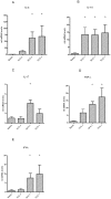
Results: 3 and 7 d after IL-1α administration, intestinal mRNA levels for IL-4, IL-10, IFN-γ and TNF-α were strongly elevated. Numbers of CD3+ and CD4+ T-lymphocytes and myeloidperoxidase+ cells were increased whereas FoxP3+ T-cells were detected at low frequency. This increased proinflammatory state was associated with ileal mucosal barrier loss as demonstrated by decreased levels of the intestinal fatty acid binding protein and disruption of the tight junctional protein ZO-1.
Conclusion: Intraamniotic IL-1α causes an acute detrimental inflammatory response in the ileum, suggesting that induction of IL-1 is a critical element in the pathophysiological effects of endotoxin induced chorioamnionitis. A disturbed balance between T-effector and FoxP3+ cells may contribute to this process.
Conflict of interest statement
Figures


Similar articles
-
Selective IL-1α exposure to the fetal gut, lung, and chorioamnion/skin causes intestinal inflammatory and developmental changes in fetal sheep.Lab Invest. 2016 Jan;96(1):69-80. doi: 10.1038/labinvest.2015.127. Epub 2015 Oct 26. Lab Invest. 2016. PMID: 26501868
-
Chorioamnionitis-induced fetal gut injury is mediated by direct gut exposure of inflammatory mediators or by lung inflammation.Am J Physiol Gastrointest Liver Physiol. 2014 Mar 1;306(5):G382-93. doi: 10.1152/ajpgi.00260.2013. Epub 2014 Jan 23. Am J Physiol Gastrointest Liver Physiol. 2014. PMID: 24458021 Free PMC article.
-
Prophylactic Interleukin-2 Treatment Prevents Fetal Gut Inflammation and Injury in an Ovine Model of Chorioamnionitis.Inflamm Bowel Dis. 2015 Sep;21(9):2026-38. doi: 10.1097/MIB.0000000000000455. Inflamm Bowel Dis. 2015. PMID: 26002542
-
Intra-amniotic IL-1β induces fetal inflammation in rhesus monkeys and alters the regulatory T cell/IL-17 balance.J Immunol. 2013 Aug 1;191(3):1102-9. doi: 10.4049/jimmunol.1300270. Epub 2013 Jun 21. J Immunol. 2013. PMID: 23794628 Free PMC article.
-
IL-1 alpha causes lung inflammation and maturation by direct effects on preterm fetal lamb lungs.Pediatr Res. 2006 Sep;60(3):294-8. doi: 10.1203/01.pdr.0000233115.51309.d3. Epub 2006 Jul 20. Pediatr Res. 2006. PMID: 16857758
Cited by
-
Bacteria and endotoxin in meconium-stained amniotic fluid at term: could intra-amniotic infection cause meconium passage?J Matern Fetal Neonatal Med. 2014 May;27(8):775-88. doi: 10.3109/14767058.2013.844124. Epub 2013 Dec 16. J Matern Fetal Neonatal Med. 2014. PMID: 24028637 Free PMC article.
-
Intraamniotic lipopolysaccharide exposure changes cell populations and structure of the ovine fetal thymus.Reprod Sci. 2013 Aug;20(8):946-56. doi: 10.1177/1933719112472742. Epub 2013 Jan 11. Reprod Sci. 2013. PMID: 23314960 Free PMC article.
-
Paneth cell ontogeny in term and preterm ovine models.Front Vet Sci. 2024 Jan 22;11:1275293. doi: 10.3389/fvets.2024.1275293. eCollection 2024. Front Vet Sci. 2024. PMID: 38318150 Free PMC article.
-
Fetal skin as a pro-inflammatory organ: Evidence from a primate model of chorioamnionitis.PLoS One. 2017 Sep 28;12(9):e0184938. doi: 10.1371/journal.pone.0184938. eCollection 2017. PLoS One. 2017. PMID: 28957335 Free PMC article.
-
Fetal immune response to chorioamnionitis.Semin Reprod Med. 2014 Jan;32(1):56-67. doi: 10.1055/s-0033-1361823. Epub 2014 Jan 3. Semin Reprod Med. 2014. PMID: 24390922 Free PMC article. Review.
References
-
- Challis JR, Lye SJ, Gibb W, Whittle W, Patel F, et al. Understanding preterm labor. Ann N Y Acad Sci. 2001;943:225–34. - PubMed
-
- Goldenberg RL, Hauth JC, Andrews WW. Intrauterine infection and preterm delivery. N Engl J Med. 2000;342:1500–7. - PubMed
-
- Andrews WW, Goldenberg RL, Faye-Petersen O, Cliver S, Goepfert AR, et al. The Alabama Preterm Birth study: polymorphonuclear and mononuclear cell placental infiltrations, other markers of inflammation, and outcomes in 23- to 32-week preterm newborn infants. Am J Obstet Gynecol. 2006;195:803–8. - PubMed
-
- Lin PW, Stoll BJ. Necrotising enterocolitis. Lancet. 2006;368:1271–83. - PubMed
-
- Sangild PT, Siggers RH, Schmidt M, Elnif J, Bjornvad CR, et al. Diet- and colonization-dependent intestinal dysfunction predisposes to necrotizing enterocolitis in preterm pigs. Gastroenterology. 2006;130:1776–92. - PubMed
Publication types
MeSH terms
Substances
Grants and funding
LinkOut - more resources
Full Text Sources
Other Literature Sources
Research Materials

