. 2011 Mar 29;6(3):e18191.
doi: 10.1371/journal.pone.0018191.

Activation of cGMP-dependent protein kinase stimulates cardiac ATP-sensitive potassium channels via a ROS/calmodulin/CaMKII signaling cascade

Affiliations

Activation of cGMP-dependent protein kinase stimulates cardiac ATP-sensitive potassium channels via a ROS/calmodulin/CaMKII signaling cascade

Yongping Chai et al. PLoS One. .

Abstract

Background: Cyclic GMP (cGMP)-dependent protein kinase (PKG) is recognized as an important signaling component in diverse cell types. PKG may influence the function of cardiac ATP-sensitive potassium (K(ATP)) channels, an ion channel critical for stress adaptation in the heart; however, the underlying mechanism remains largely unknown. The present study was designed to address this issue.

Methods and findings: Single-channel recordings of cardiac K(ATP) channels were performed in both cell-attached and inside-out patch configurations using transfected human embryonic kidney (HEK)293 cells and rabbit ventricular cardiomyocytes. We found that Kir6.2/SUR2A (the cardiac-type K(ATP)) channels were activated by cGMP-selective phosphodiesterase inhibitor zaprinast in a concentration-dependent manner in cell-attached patches obtained from HEK293 cells, an effect mimicked by the membrane-permeable cGMP analog 8-bromo-cGMP whereas abolished by selective PKG inhibitors. Intriguingly, direct application of PKG moderately reduced rather than augmented Kir6.2/SUR2A single-channel currents in excised, inside-out patches. Moreover, PKG stimulation of Kir6.2/SUR2A channels in intact cells was abrogated by ROS/H(2)O(2) scavenging, antagonism of calmodulin, and blockade of calcium/calmodulin-dependent protein kinase II (CaMKII), respectively. Exogenous H(2)O(2) also concentration-dependently stimulated Kir6.2/SUR2A channels in intact cells, and its effect was prevented by inhibition of calmodulin or CaMKII. PKG stimulation of K(ATP) channels was confirmed in intact ventricular cardiomyocytes, which was ROS- and CaMKII-dependent. Kinetically, PKG appeared to stimulate these channels by destabilizing the longest closed state while stabilizing the long open state and facilitating opening transitions.

Conclusion: The present study provides novel evidence that PKG exerts dual regulation of cardiac K(ATP) channels, including marked stimulation resulting from intracellular signaling mediated by ROS (H(2)O(2) in particular), calmodulin and CaMKII, alongside of moderate channel suppression likely mediated by direct PKG phosphorylation of the channel or some closely associated proteins. The novel cGMP/PKG/ROS/calmodulin/CaMKII signaling pathway may regulate cardiomyocyte excitability by opening K(ATP) channels and contribute to cardiac protection against ischemia-reperfusion injury.


Conflict of interest statement

Competing Interests: The authors have declared that no competing interests exist.

Figures

Figure 1
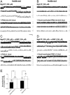
Figure 1. Stimulation of Kir6.2/SUR2A channels by activation of PKG in transfected HEK293 cells.
Recombinant Kir6.2/SUR2A channels were expressed in HEK293 cells by transient transfection. Recordings were performed in symmetrical high potassium (140-mM) solutions at room temperature in the cell-attached configuration and the membrane potential was clamped at −60 mV. (A–C) Single-channel current traces of the Kir6.2/SUR2A channel obtained from a representative cell-attached patch prior to (upper panel) and during (lower panel) application of the membrane-permeable cGMP analog 8-Br-cGMP (500 µM) (A), the cGMP-specific PDE inhibitor zaprinast (50 µM) alone (B), or zaprinast (50 µM) plus a selective PKG inhibitor KT5823 (1 µM) following pretreatment with KT5823 for 15 min (C). Downward deflections represent openings from closed states. For all current trace figures, segments of raw recordings marked with a horizontal line on top are shown in successive traces at increasing temporal resolution, revealing singular openings (*) and bursts of openings (**). The horizontal scale bars represent 1 s, 300 ms and 100 ms for traces from top to bottom in each three-trace panel, and the vertical scale bar represents 4 pA. (D) The averaged normalized open probability NPo (i.e., relative channel activity) of Kir6.2/SUR2A channels obtained during application of drugs in individual groups of cell-attached patches. Data obtained from patches treated with increasing concentrations of zaprinast (0.05, 0.5, 5 and 50 µM) were also displayed to illustrate the concentration dependence of zaprinast effects. NPo values of all groups were normalized to the corresponding control (taken as 1; dashed line) obtained prior to index drug application in individual patches to yield the normalized NPo. Data are presented as mean ± SEM of 6–12 patches (number of patches in individual groups provided in parentheses). Significance levels are: *, P<0.05; **, P<0.01; ***, P<0.001 (two-tailed one-sample t tests within individual groups, or unpaired t tests between groups).
Figure 2
Figure 2. Suppression of Kir6.2/SUR2A channel activity by direct application of purified PKG in excised inside-out patches.
Single-channel currents were obtained in the inside-out patch configuration from transiently transfected HEK293 cells. Recordings were conducted in symmetrical high potassium (140-mM) solutions and the membrane potential was clamped at −60 mV. (A–D) Single-channel current traces of Kir6.2/SUR2A channels in a representative inside-out patch prior to (upper panel) and during (lower panel) bath perfusion of the catalytic subunit of PKG (PKG-CA; 0.5 U/µl) (A), heat-inactivated PKG-CA (B), purified PKG Iα holoenzyme (PKG; 0.5 U/µl) (C), or heat-inactivated PKG holoenzyme (D). Numbers (from 0–2) marked at the right margin of current traces indicate the level of simultaneous channel opening: 0 for closed-channel, 1 for one-channel, and 2 for two-channel level opening. Scale bars are the same as described in Fig. 1. The single-channel currents of KATP channels in inside-out patches were usually higher than in cell-attached patches (Figs. 2A vs. 1A), as ATP inhibition of the channel was partially alleviated upon patch excision. (E) Normalized NPo values of Kir6.2/SUR2A channels obtained during applications of live or heat-inactivated PKG in individual groups of inside-out patches. The drug effect was compared with the corresponding control obtained from the same patch and NPo values were normalized as described in Fig. 1D (control taken as 1; dashed line). The average data are presented as mean ± SEM of 5–8 patches. Significance level is: *, P<0.05; **, P<0.01 (two-tailed one-sample t tests within individual groups, or unpaired t tests between groups).
Figure 3
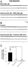
Figure 3. The role of ROS, particularly H2O2, in mediating Kir6.2/SUR2A channel stimulation downstream of PKG activation.
Single-channel currents were obtained from cell-attached patches obtained from transiently transfected HEK293 cells. Recordings and drug application were administered as described in Fig. 1. (A–B) Single-channel current traces of the Kir6.2/SUR2A channel obtained from a cell-attached patch prior to (upper panel) and during (lower panel) application of the cGMP-specific PDE inhibitor zaprinast (50 µM) together with a membrane-permeable ROS scavenger MPG (500 µM) after 15-min preincubation in MPG (A), or with catalase, a H2O2-decomposing enzyme (500 U/ml) (B). There was no pretreatment for cells in the catalase group because catalase is not membrane-permeable. Scale bars are the same as described in Fig. 1. (C) Normalized NPo values of Kir6.2/SUR2A channels obtained during applications in individual groups of cell-attached patches. NPo values were normalized to the corresponding control as described in Fig. 1D (control taken as 1; dashed line). The zaprinast data (filled bar) are the same as presented in Fig. 1D, and are included here for comparison purpose. Data are presented as mean ± SEM of 6–12 patches. The significance level is: **, P<0.01 (two-tailed one-sample t tests within individual groups, or Dunnett's multiple comparison tests between groups).
Figure 4
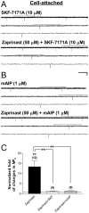
Figure 4. Roles of calmodulin and CaMKII in mediating PKG stimulation of Kir6.2/SUR2A channels.
Recombinant Kir6.2/SUR2A channels were expressed in HEK293 cells by transient transfection. Cell-attached patch-clamp recordings and drug application were administered as described in Fig. 1. (A–B) Single-channel current traces of Kir6.2/SUR2A channel obtained from a cell-attached patch prior to (upper panel) and during (lower panel) application of the cGMP-specific PDE inhibitor zaprinast (50 µM) together with the membrane-permeable, selective calmodulin antagonist SKF-7171A (10 µM) (A), or with the myristoylated autocamtide-2 related inhibitory peptide for CaMKII (mAIP; 1 µM) (B). The scale bars are the same as described in Fig. 1. (C) Normalized NPo of Kir6.2/SUR2A channels obtained during application of drugs in individual groups. The zaprinast data (filled bar) are the same as presented in Fig. 1D, and are included here for comparison purpose. NPo values were normalized to the corresponding control (taken as 1) as described in Fig. 1D. The dashed line indicates the control level. Data are presented as mean ± SEM of 8–12 patches. The significance level is: **, P<0.01 (two-tailed one-sample t tests within individual groups, or Dunnett's multiple comparison tests between groups).
Figure 5
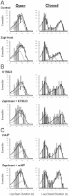
Figure 5. Effects of PKG activation on open- and closed-duration distributions of Kir6.2/SUR2A channels in intact cells.
Data were obtained from transfected HEK293 cells expressing Kir6.2/SUR2A channels. Frequency histograms of duration distributions fitted from events obtained before (upper panel) and during (lower panel) the application of zaprinast (50 µM) alone (A), zaprinast (50 µM) plus a selective PKG inhibitor KT5823 (1 µM) (B), or zaprinast (50 µM) plus the myristoylated CaMKII inhibitory peptide mAIP (1 µM) (C) in representative cell-attached patches. Cells were incubated with respective inhibitors for at least 15 min before index drug perfusion. Frequency histograms displayed are open-duration distribution (left column) and closed-duration distributions (right column), respectively, for all patches. Duration histograms were constructed as described in Methods.
Figure 6
Figure 6. Dual effects of H2O2 on the function of cardiac-type KATP channels.
Currents were obtained in the cell-attached (A,B) or inside-out (C) patch configuration from transiently transfected HEK293 cells expressing Kir6.2/SUR2A channels. (A–B) Single-channel current traces of Kir6.2/SUR2A channels in representative cell-attached patches prior to (upper panel) and during (lower panel) bath perfusion of H2O2 (1 mM) alone (A), or H2O2 plus the myristoylated CaMKII inhibitory peptide mAIP (1 µM) (B). (C) Single-channel current traces of Kir6.2/SUR2A channels from a representative inside-out patch before and during application of H2O2 (1 mM). Numbers (from 0–4) provided along the right margin of current traces indicate the increasing level of simultaneous channel opening: 0 for closed-channel, 1 for one-channel, 2 for two-channel level opening, etc. MgATP (30 µM) was included in the bath and drug solutions during inside-out recordings to prevent current rundown. The scale bars are the same as described in Fig. 1. (D) Normalized NPo of Kir6.2/SUR2A channels obtained during application of drugs in individual groups. The labels (cell-attached and inside-out) placed underneath the X axis depict the patch configuration in which data were obtained. NPo values of all groups were normalized to the corresponding control obtained prior to index drug application in individual patches as described in Fig. 1D (control taken as 1; dashed line). Data obtained from cell-attached patches treated with increasing concentrations of H2O2 (0.0001, 0.01 and 1 mM) were also displayed (first three bars from the left) to illustrate the concentration dependence of H2O2 effects. Data are presented as mean ± SEM of 3–11 patches (number of patches in individual groups provided in parentheses). Significance levels are: *, P<0.05; **, P<0.01 (two-tailed one-sample t tests within individual groups, or unpaired t tests between groups).
Figure 7
Figure 7. Stimulation of sarcKATP channels in ventricular cardiomyocyte by intracellular signaling triggered by activation of PKG.
Recordings were performed in symmetrical high potassium (140-mM) solutions at room temperature in the cell-attached configuration and the membrane potential was clamped at −60 mV. (A–D) Single-channel current traces of the sarcKATP channel preactivated by the KATP channel opener pinacidil (200 µM) in a representative cell-attached patch prior to (upper panel) and during (lower panel) addition of the cGMP-specific PDE inhibitor zaprinast (50 µM) (A), or zaprinast (50 µM) together with one of the following: a selective PKG inhibitor KT5823 (1 µM) (B), a membrane-permeable ROS scavenger MPG (500 µM) (C), or the myristoylated autocamtide-2 related inhibitory peptide for CaMKII (mAIP; 1 µM) (D). Cells were pretreated with respective inhibitors for at least 15 min at room temperature before recordings were started. Scale bars are the same as described in Fig. 1. Downward deflections represent openings from closed states. (E) The averaged normalized NPo values of sarcKATP channels obtained during application of drugs in individual groups of cell-attached patches. NPo values were normalized to the corresponding control in pinacidil with or without inhibitors (control taken as 1; dashed line). Data are presented as mean ± SEM of 4–9 patches. Significance levels are: *, P<0.05; **, P<0.01 (two-tailed one-sample t tests within individual groups, or Dunnett's multiple comparison tests between groups).

Similar articles

Cited by

References

    1. Ashcroft FM. Adenosine 5′-triphosphate-sensitive potassium channels. Annu Rev Neurosci. 1988;11:97–118. - PubMed
    1. Miki T, Seino S. Roles of KATP channels as metabolic sensors in acute metabolic changes. J Mol Cell Cardiol. 2005;38:917–925. - PubMed
    1. Nichols CG. KATP channels as molecular sensors of cellular metabolism. Nature. 2006;440:470–476. - PubMed
    1. Shyng S-L, Nichols CG. Octameric stoichiometry of KATP channel complex. J Gen Physiol. 1997;110:655–664. - PMC - PubMed
    1. Babenko AP, Aguilar-Bryan L, Bryan J. A view of SUR/Kir6.X, KATP channels. Annu Rev Physiol. 1998;60:667–687. - PubMed

Publication types

MeSH terms