Review
. 2012 Jan;41(1):15-31.
doi: 10.1007/s00256-011-1143-1. Epub 2011 Apr 10.

High resolution imaging of tunnels by magnetic resonance neurography

Affiliations
Review

High resolution imaging of tunnels by magnetic resonance neurography

Ty K Subhawong et al. Skeletal Radiol. 2012 Jan.

Abstract

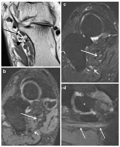
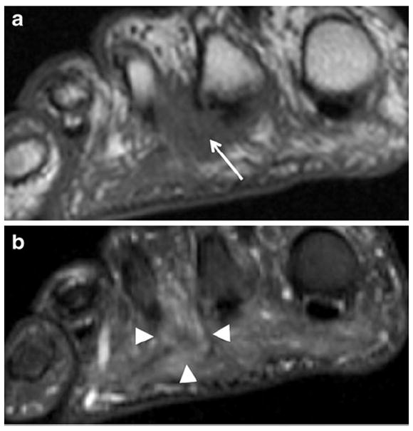
Peripheral nerves often traverse confined fibro-osseous and fibro-muscular tunnels in the extremities, where they are particularly vulnerable to entrapment and compressive neuropathy. This gives rise to various tunnel syndromes, characterized by distinct patterns of muscular weakness and sensory deficits. This article focuses on several upper and lower extremity tunnels, in which direct visualization of the normal and abnormal nerve in question is possible with high resolution 3T MR neurography (MRN). MRN can also serve as a useful adjunct to clinical and electrophysiologic exams by discriminating adhesive lesions (perineural scar) from compressive lesions (such as tumor, ganglion, hypertrophic callous, or anomalous muscles) responsible for symptoms, thereby guiding appropriate treatment.


Conflict of interest statement

Conflict of interest The authors report no conflicts of interest.

Figures

Fig. 1
Fig. 1
A 14-year-old boy with wrist pain. a Axial T1 image shows the normal fascicular pattern of the median nerve in the carpal tunnel (arrow), with well-preserved planes of surrounding perineural fat. b Axial T2 SPAIR image shows the normal intermediate signal intensity of the median nerve (white arrow), in contrast to the hyperintense vessels (black arrow)
Fig. 2
Fig. 2
Sagittal PSIF (nerve selective) image demonstrates the normal course of the median nerve as it travels ventrally in the forearm
Fig. 3
Fig. 3
Coronal 3-D maximum intensity projection (MIP) from a STIR SPACE sequence of the brachial plexus demonstrates improved conspicuity of the nerve roots (arrows)
Fig. 4
Fig. 4
A 60-year-old man with left wrist pain due to carpal tunnel syndrome (idiopathic). Axial T2 SPAIR shows significant T2 hyperintensity and enlargement of the median nerve as well as contained fascicles (arrow), with minimal flexor tenosynovitis and volar bowing of the overlying flexor retinaculum (arrowheads)
Fig. 5
Fig. 5
A 25-year-old man with history of trauma to right thumb and persistent pain. Axial T2 SPAIR images show a persistent median artery (white arrow) between the abnormally T2 hyperintense radial and ulnar bundles of the bifid median nerve (black arrows). A persistent median artery is a known cause of compression median neuropathy in the carpal tunnel
Fig. 6
Fig. 6
A 31-year-old male with ulnar sided wrist pain. Axial T2 SPAIR image just distal to Guyon’s canal shows an isolated signal abnormality in the deep branch of the ulnar nerve (white arrow); note the associated denervation edema of the hypothenar muscles (black arrow). The more superficial branches are normal (arrowhead)
Fig. 7
Fig. 7
A 45-year-old man with history of forearm pain. a Axial T1 and b axial T2 SPAIR images show an accessory anconeus muscle (black arrow) causing compression and ulnar neuropathy (white arrow)
Fig. 8
Fig. 8
A 51-year-old male with persistent left upper arm pain as well as left hand pain and numbness, following prior surgical ulnar nerve transposition. a Axial T1 image demonstrates anterior subcutaneous position of the ulnar nerve (arrow). Note thin strand hypointense fibrotic bands (arrowhead) surrounding the nerve. b Axial T2 SPAIR shows abnormal hyperintensity of the ulnar nerve (arrow), approaching that of vessels. c Photograph from surgery shows the fibrous bands around the inflamed and hyperemic ulnar nerve (arrow), confirming the pre-operative MRI findings
Fig. 9
Fig. 9
A 45-year-old man with elbow pain and weakness in finger extension. a Oblique coronal STIR MIP shows the abnormal radial nerve (long arrow), which is enlarged and hyperintense related to hypothesized mechanisms of blocked axoplasmic flow and venous congestion proximal to the site of entrapment at the arcade of Frohse (short arrow). b Axial T2 SPAIR image demonstrates the abnormal radial nerve, which shows abnormal hyperintense signal in the T2 weighted image (white arrow). c Axial T2 SPAIR more distally in the forearm shows extensive denervation edema-like signal affecting extensor compartment muscles (black arrow). MRI confirmed the clinical suspicion of posterior interosseous neuropathy and excluded the presence of any organic cause. The symptoms resolved on clinical follow-up
Fig. 10
Fig. 10
A 54-year-old with right thumb weakness and clinically suspected AIN neuropathy. a Axial T2 SPAIR shows high signal intensity (similar to vessels) of the AIN (black arrow). Axial T2 SPAIR images show denervation edema-like signal of the flexor digitorum profundus muscle (b, thick white arrow) as well as of the pronator quadratus muscle (c, thin white arrow)
Fig. 11
Fig. 11
A 50-year-old man with shoulder weakness. Sagittal STIR image shows an abnormally hyperintense suprascapular nerve (encircled) entrapped at the suprascapular notch, with consequent subacute denervation edema of the supra- and infraspinatus muscles (arrows). The patient successfully responded to the suprascapular ligament release. No mass lesion was found intraoperatively
Fig. 12
Fig. 12
A 24-year-old man with shoulder pain and weakness. Coronal proton-density weighted (PDW) image with fat saturation shows a SLAP type II tear (arrow), with paralabral cyst formation in the spinoglenoid notch (arrowhead), causing entrapment of the supra-scapular nerve and resultant denervation changes of the infraspinatus muscle (oval)
Fig. 13
Fig. 13
A 71-year-old woman with shoulder pain. a Sagittal PD images show large humeral osteophytes (arrow) projecting into the quadrilateral space. b Sagittal PD image medial to a shows selective denervation atrophy of the teres minor muscle (oval)
Fig. 14
Fig. 14
A 35-year-old woman with left arm pain. a Coronal T1 of the thoracic outlet shows narrowing of the interscalene compartment by a prominent transverse process of C7 (black arrow). b Sagittal STIR image shows abnormal enlargement and T2 hyperintensity of the C8 and T1 nerve roots (arrow), consistent with thoracic outlet syndrome
Fig. 15
Fig. 15
A 31-year-old woman with calf pain. a Axial T2 SPAIR image shows normal tibial nerve (arrow) proximal to the soleal sling. b Axial T2 SPAIR image (more distal than a) demonstrates subtle hyperintensity of the tibial nerve (long arrow) as it crosses under the sling (not well visualized); note normal signal intensity of the common peroneal nerve (short arrow). After surgical release of the entrapment, the patient’s symptoms improved
Fig. 16
Fig. 16
A 44-year-old male with left foot drop after sinus surgery, and EMG, which showed evidence of left peroneal neuropathy at the fibular head. a Axial T2 SPAIR demonstrates increased signal intensity of the peroneal nerve (arrow) posterior to the fibular head in the peroneal tunnel area as well as denervation edema-like signal in the anterior compartment muscles (encircled). b Axial T2 SPAIR (inferior to a) shows grade 1 strain of the lateral head of the gastrocnemius muscle (encircled), suggesting local compression due to inattention to proper patient positioning during surgery as the cause of injury
Fig. 17
Fig. 17
A 58-year-old woman with previous tarsal tunnel release and persistent ankle pain in medial plantar nerve distribution. a Axial T1 FLAIR demonstrates the medial (long arrow) and lateral (short arrow) plantar nerves in their respective tunnels; note the thin fibrous septum (arrowhead) between the tunnels. b Coronal oblique PSIF and c SPAIR show subtle increased hyperintensity of the MPN (long white arrow) as well as hyperintensity of the LPN (short white arrow). d Sagittal PSIF MIP shows enlargement of the entrapped MPN (arrows); note that vasculature adjacent to the nerves is suppressed on the PSIF images. For orientation, an asterisk demarks the talus
Fig. 18
Fig. 18
A 58-year-old woman with foot pain. Coronal (short-axis axial) T1 image through the distal metatarsals shows intermediate signal intensity mass in the 2nd intermetatarsal web space (arrow) with effacement of the fat planes around the common digital nerve. Coronal (short-axis axial) STIR image shows mild hyperintensity within this Morton’s neuroma (arrowheads)

Similar articles

Cited by

References

    1. Siemionow M, Brzezicki G. Chapter 8: current techniques and concepts in peripheral nerve repair. Int Rev Neurobiol. 2009;87:141–72. - PubMed
    1. Pećina M, Krmpotić-Nemanić J, Markiewitz AD. Tunnel syndromes. 3. Boca Raton: CRC Press; 2001.
    1. Gutmann L. Pearls and pitfalls in the use of electromyography and nerve conduction studies. Semin Neurol. 2003;23:77–82. - PubMed
    1. Bashir WA, Connell DA. Imaging of entrapment and compressive neuropathies. Semin Musculoskelet Radiol. 2008;12:170–81. - PubMed
    1. Spratt JD, Stanley AJ, Grainger AJ, Hide IG, Campbell RSD. The role of diagnostic radiology in compressive and entrapment neuropathies. Eur Radiol. 2002;12:2352–64. - PubMed

Publication types