. 2011 Dec;68(23):3885-901.
doi: 10.1007/s00018-011-0679-5. Epub 2011 Apr 10.

Roles of the human hypoxia-inducible factor (HIF)-3α variants in the hypoxia response

Affiliations

Roles of the human hypoxia-inducible factor (HIF)-3α variants in the hypoxia response

Minna Heikkilä et al. Cell Mol Life Sci. 2011 Dec.

Abstract

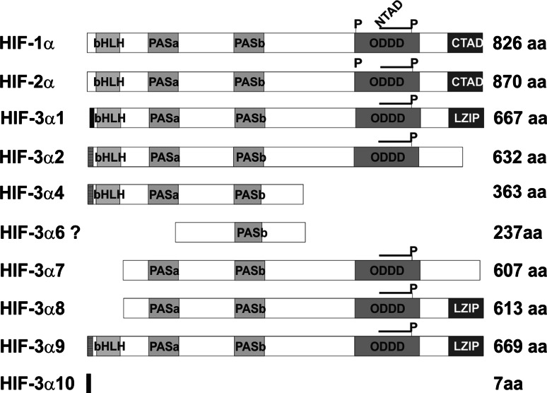
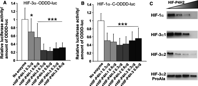
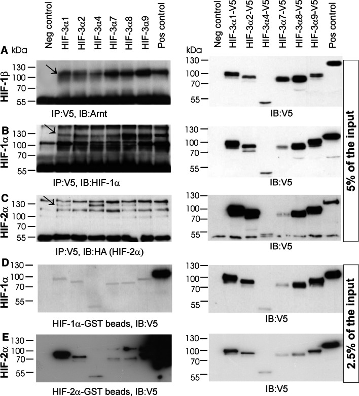
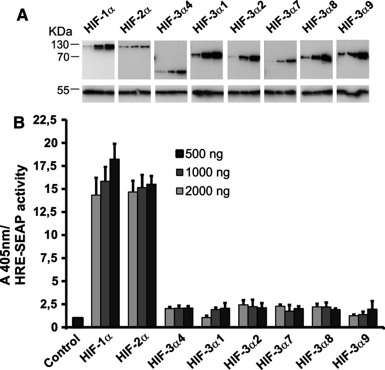
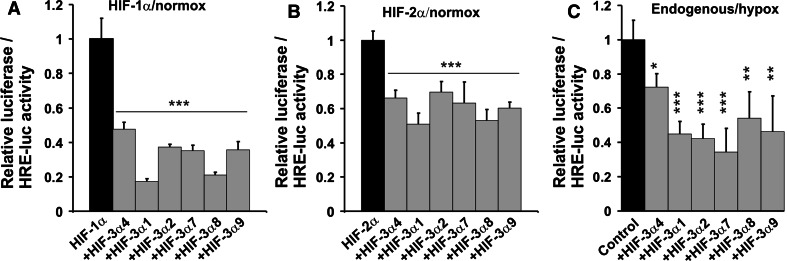
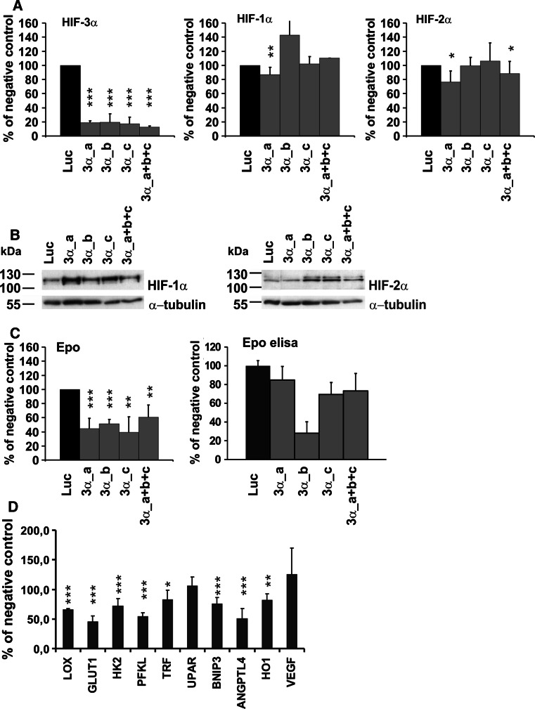
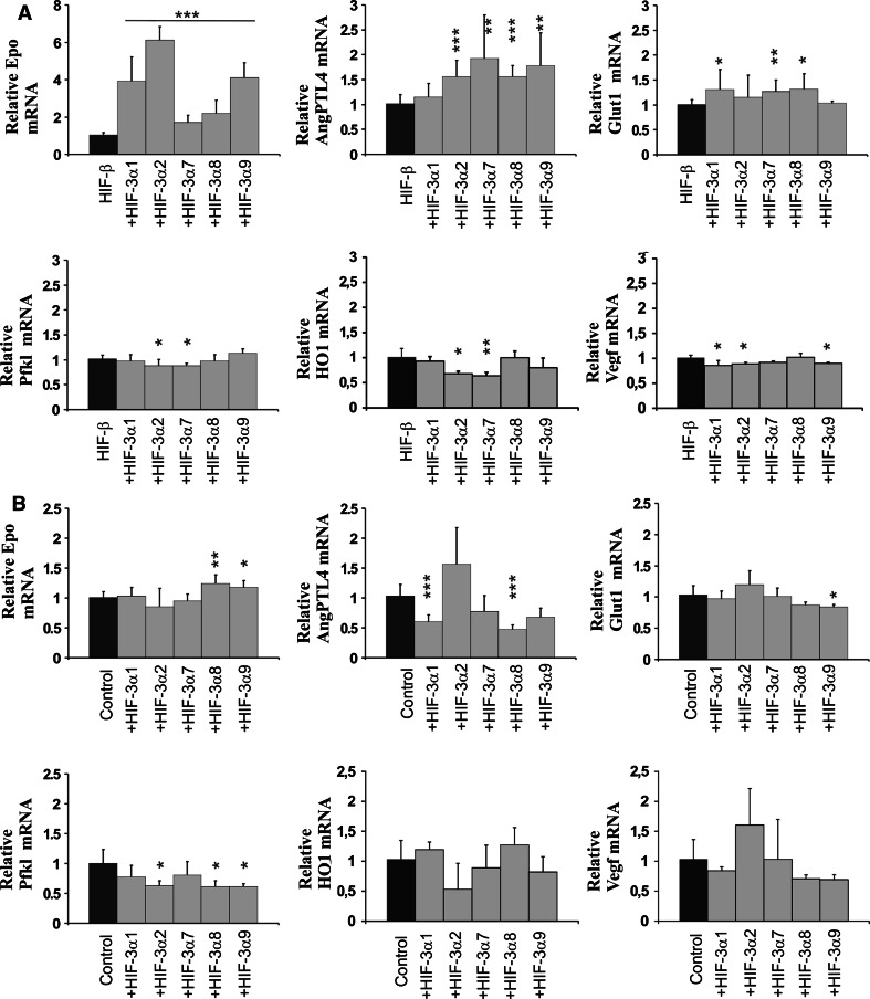
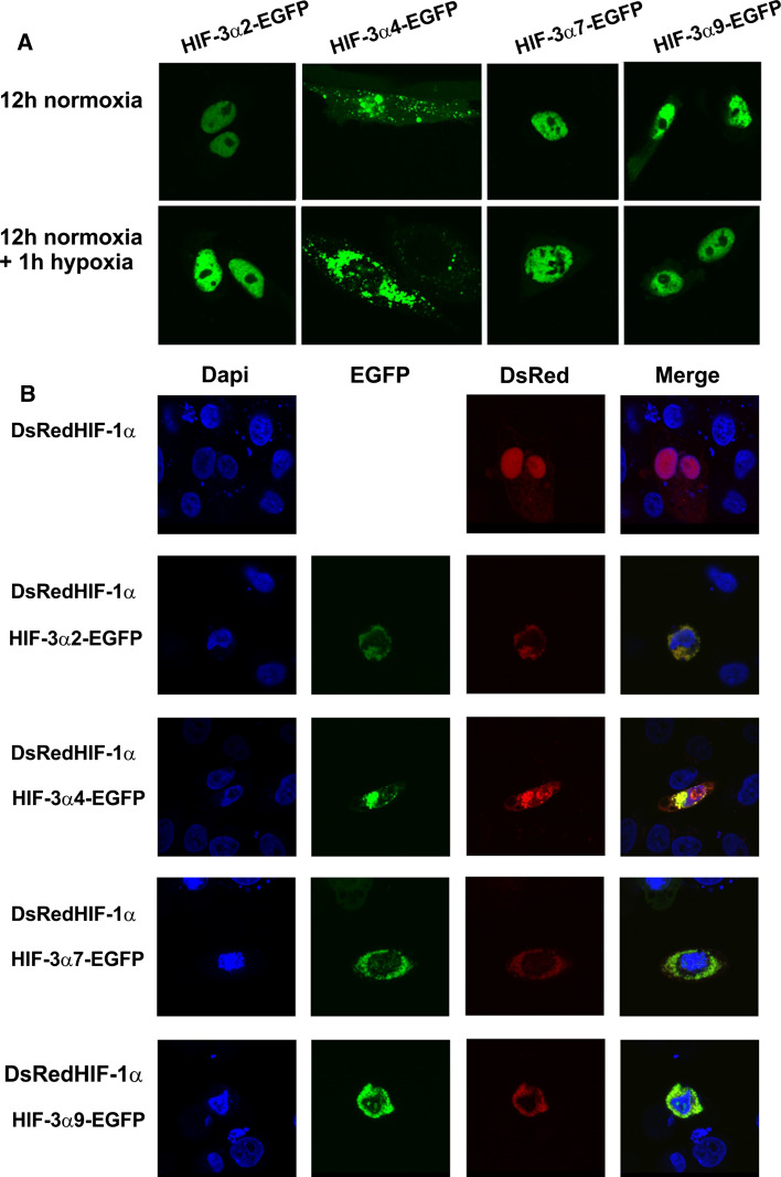
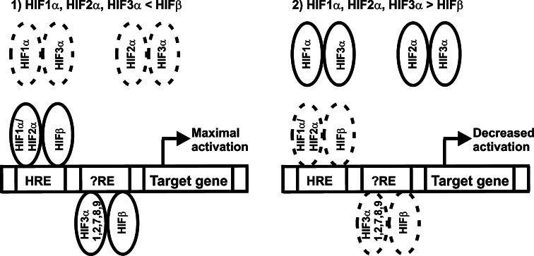
The hypoxia-inducible transcription factor (HIF) controls (in an oxygen-dependent manner) the expression of a large number of genes whose products are involved in the response of cells to hypoxia. HIF is an αβ dimer that binds to hypoxia response elements (HREs) in its target genes. Human HIF-α has three isoforms, HIF-1α, HIF-2α and HIF-3α, of which the roles of HIF-3α are largely unknown, although it is usually regarded as a negative regulator of HIF-1α and HIF-2α. The human HIF-3α locus is subject to extensive alternative splicing, leading to at least seven variants. We analyzed here the effects of the long variants and the short variant HIF-3α4 on the hypoxia response. All these variants were found to interact with HIF-β, HIF-1α and HIF-2α. The long HIF-3α variants were localized in the nucleus in hypoxia, while HIF-3α4 was cytoplasmic. Interaction of the HIF-3α variants with HIF-1α inhibited the nuclear translocation of both. None of the long HIF-3α variants was capable of efficient induction of an HRE reporter in overexpression experiments, but instead inhibited the transcriptional activation of the reporter by HIF-1 and HIF-2. Unexpectedly, siRNA knock-down of the endogenous HIF-3α variants led to downregulation of certain HIF target genes, while overexpression of individual long HIF-3α variants upregulated certain HIF target genes in a variant and target gene-specific manner under conditions in which HIF-β was not a limiting factor. These data indicate that the HIF-3α variants may have more versatile and specific roles in the regulation of the hypoxia response than previously anticipated.


Figures

Fig. 1
Fig. 1
Schematic representation of the HIF-1α, HIF-2α, and the HIF-3α variant polypeptides. The structural motifs basic helix-loop-helix (bHLH), Per/Arnt/Sim (PAS), the oxygen-dependent degradation domain (ODDD), the N and C-terminal transactivation domains (NTAD and CTAD), the leucine zipper (LZIP), and the lengths of the polypeptides are indicated. The positions of the prolines hydroxylated by HIF-P4Hs in the ODDD are indicated by P
Fig. 2
Fig. 2
Overexpression of HIF-P4H enzymes leads to degradation of a HIF-3α-ODDD reporter and full-length HIF-3 variants. ChoK1 cells were transfected with a HIF-3α-ODDD-luc (a) or HIF-1α-C-ODDD-luc (b) reporter plasmid together with 0.3 or 0.6 μg of HIF-P4H-1, HIF-P4H-2 or HIF-P4H-3 expression plasmids. The transfected cells were cultured for 24 h in normoxia and assayed for luciferase activity. The luciferase activity of the control sample without HIF-P4H overexpression was set as 1. The data represent means ± SD from at least four independent experiments. *p < 0.05, ***p < 0.001. To study the degradation of full-length HIF-α polypeptides, ChoK1 cells were transfected with plasmids encoding full-length HIF-1α, V5-tagged HIF-3α1, HIF-3α2 or HIF-3α2_ProAla together with increasing amounts of HIF-P4H-2 plasmid (c). The cells were cultured for 48 h in normoxia, lysed, and analyzed by 8% SDS-PAGE under reducing conditions followed by Western blotting with HIF-1α, V5 and α-tubulin antibodies. The results of a typical experiment are shown
Fig. 3
Fig. 3
All HIF-3α variants can bind HIF-1α, HIF-2α and HIF-1β. ChoK1 cells were transfected for immunoprecipitation studies with plasmids for V5-tagged HIF-3α1, HIF-3α2, HIF-3α4, HIF-3α7, HIF-3α8 and HIF-3α9 with or without untagged HIF-1β (a), HIF-1α (b), or HA-tagged HIF-2α (c). In the positive control samples the cells were transfected with plasmids for HIF-1β and V5-tagged HIF1α (a), HIF-1α and V5-tagged HIF-β (b), and HA-tagged HIF-2α andV5-tagged HIF-β (c). The cells were cultured for 24 h in normoxia followed by 24 h in 1% oxygen. The left-hand panels in a–c show the amounts of the HIF-1β, HIF-1α and HIF-2α polypeptides immunoprecipitated with the V5-tagged HIF-3α variants or the positive controls, and the right-hand panels show the amounts of the V5-tagged polypeptides present in the whole cell lysates (5% of the input amount) used in the immunoprecipitation analysis (d–e). For in vitro binding studies HIF-1α and HIF-2α were expressed as GST-fusion proteins in E. coli, purified and bound to glutathione agarose beads. ChoK1 cells were transfected with plasmids for the V5-tagged HIF-3α variants or HIF-β (positive control) or the empty vector pcDNA3.1/Zeo(−) (negative control) and cultured for 24 h in normoxia followed by 24 h in 1% oxygen. The cell lysates were incubated with the GST beads ± GST-HIF-1α or GST-HIF-2α. The left-hand panels show the amounts of the captured V5-tagged HIF-3α variants or positive controls, and the right-hand panels show the amounts of the V5-tagged polypeptides present in the whole cell lysates (2.5% of the input amount) used in the in vitro binding assay
Fig. 4
Fig. 4
Effects of the HIF-3α variants on the activation of a HRE reporter. ChoK1 cells were transfected with an HRE-SEAP reporter plasmid together with increasing amounts (500, 1,000, 2,000 ng) of expression plasmids for HIF-1α, HIF-2α or the HIF-3α variants. The cells were cultured for 48 h in normoxia and analyzed for expression of the HIF-α polypeptides by Western blotting (a) and assayed for SEAP activity (b), which was set to 1 in the control sample without any overexpressed HIF-α polypeptide. The results of a typical experiment are shown in a, and the data in b represent means ± S.D. from at least 4 independent experiments
Fig. 5
Fig. 5
HIF-3α variants inhibit the transcriptional activation of an HRE-reporter by HIF-1 and HIF-2. ChoK1 cells were transfected with a 3xHRE-LUC reporter plasmid together with plasmids for HIF-1α (a) or HIF-2α (b) and with or without plasmids for the HIF-3α variants. The cells were cultured for 48 h in normoxia and assayed for luciferase activity. The luciferase activity of cells transfected only with HIF-1α or HIF-2α was set to 1. The data represent means ± SD from at least six independent experiments. ***p < 0.001. Hep3B cells were transfected with a 3xHRE-Luc reporter plasmid together with a empty vector or certain HIF-3α variants (c). Transfected cells were cultured for 24 h in normoxia followed by 24 h in hypoxia and then assayed for luciferase activity. The luciferase activity of the control samples was set to 1
Fig. 6
Fig. 6
siRNA knock-down of endogenous HIF-3α leads to downregulation of many HIF target genes. Hep3B cells were transfected with a negative control siRNA (luc, pGL2 luciferase control duplex siRNA) or with HIF-3α siRNAs (three independent siRNAs a, b, c alone or in combination) twice at an interval of 24 h and cultured in 1% oxygen for 24 h after the second transfection. The expression of HIF-3α (all variants), HIF-1α, HIF-2α (a) and Epo (c) at the mRNA level was analyzed by qPCR and the expression of HIF-1α and HIF-2α (b) and Epo (c) at the protein level by Western blotting and ELISA, respectively. The effect of the HIF-3α siRNA on selected HIF target genes was analyzed by qPCR (d). The data represent means ± SD from at least four independent experiments. *p < 0.05, **p < 0.01, ***p < 0.001. In the case of the Epo ELISA analysis, the data represent the results of a typical experiment
Fig. 7
Fig. 7
Certain long HIF-3α variants upregulate HIF target genes when HIF-β is not limiting. Hep3B cells were transfected with a plasmid encoding the full-length long HIF-3α variants with (a) or without (b) HIF-β. HIF-3α4 was excluded here as the aim was to study the transactivation potential of HIF-3α variants. The cells were cultured for 48 h in normoxia followed by 90 min in 1% oxygen (a) or for 24 h in normoxia followed by 24 h in 1% oxygen (b). The expression levels of the EPO, ANGPTL4, GLUT1, PFKL, HO-1 and VEGF mRNAs were analyzed by qPCR. The data represent means ± SD from at least three independent experiments. *p < 0.05, **p < 0.01,***p < 0.001
Fig. 8
Fig. 8
Confocal microscopy analysis of the cellular localization of overexpressed HIF-3α variants. a ChoK1 cells were transfected with plasmids encoding EGFP-labeled HIF-3α variants 2, 4, 7, or 9 and cultured in normoxia for 12 h (upper panel) followed by 1 h in 1% oxygen (lower panel). b ChoK1 cells were cotransfected with plasmids encoding EGFP-labeled HIF-3α variants 2, 4, 7, or 9 and DSRed-labeled HIF-1α and cultured for 24 h in normoxia followed by 8 h in 1% oxygen and stained with DAPI after fixation
Fig. 9
Fig. 9
Schematic representation of the roles of long HIF-3α variants in the hypoxia response. a When HIF-β is not limiting, HIF-1α, HIF-2α, and the long HIF-3α variants mainly associate with HIF-β and activate hypoxia-inducible target genes. Our data suggest that the long HIF-3α variants are required for maximal activation of certain HIF target genes and are likely to have target elements distinct from the canonical HRE response element. b When the amount of HIF-β is limiting, the long HIF-3α variants associate with HIF-1α and HIF-2α leading to decreased activation of HIF target genes. The short variant HIF-3α4, which is not depicted in the scheme, forms inactive complexes with HIF-1α, HIF-2α and HIF-β and causes downregulation of HIF target genes independent on whether HIF-β is non-limiting or limiting. Formation of these complexes is also dependent on the relative amounts of HIF-3α4, HIF-1α, HIF-2α and HIF-β

References

    1. Semenza GL. HIF-1: upstream and downstream of cancer metabolism. Curr Opin Genet Dev. 2010;20:51–56. doi: 10.1016/j.gde.2009.10.009. - DOI - PMC - PubMed
    1. Kaelin WG, Jr, Ratcliffe PJ. Oxygen sensing by metazoans: the central role of the HIF hydroxylase pathway. Mol Cell. 2008;30:393–402. doi: 10.1016/j.molcel.2008.04.009. - DOI - PubMed
    1. Weidemann A, Johnson RS. Biology of HIF-1α. Cell Death Differ. 2008;15:621–627. doi: 10.1038/cdd.2008.12. - DOI - PubMed
    1. Myllyharju J. Prolyl 4-hydroxylases, key enzymes in the synthesis of collagens and regulation of the response to hypoxia, and their roles as treatment targets. Ann Med. 2008;40:402–417. doi: 10.1080/07853890801986594. - DOI - PubMed
    1. Ivan M, Kondo K, Yang H, Kim W, Valiando J, Ohh M, Salic A, Asara JM, Lanie WS, Kaelin WG., Jr HIFα targeted for VHL-mediated destruction by proline hydroxylation: implications for O2 sensing. Science. 2001;292:464–468. doi: 10.1126/science.1059817. - DOI - PubMed

Publication types

MeSH terms

Substances

LinkOut - more resources