. 2011 Jul;401(1):167-81.
doi: 10.1007/s00216-011-4929-z. Epub 2011 Apr 12.

Normalization in MALDI-TOF imaging datasets of proteins: practical considerations

Affiliations

Normalization in MALDI-TOF imaging datasets of proteins: practical considerations

Sören-Oliver Deininger et al. Anal Bioanal Chem. 2011 Jul.

Abstract

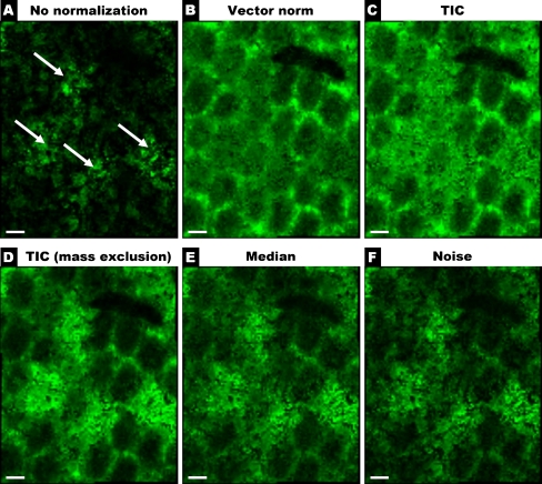
Normalization is critically important for the proper interpretation of matrix-assisted laser desorption/ionization (MALDI) imaging datasets. The effects of the commonly used normalization techniques based on total ion count (TIC) or vector norm normalization are significant, and they are frequently beneficial. In certain cases, however, these normalization algorithms may produce misleading results and possibly lead to wrong conclusions, e.g. regarding to potential biomarker distributions. This is typical for tissues in which signals of prominent abundance are present in confined areas, such as insulin in the pancreas or β-amyloid peptides in the brain. In this work, we investigated whether normalization can be improved if dominant signals are excluded from the calculation. Because manual interaction with the data (e.g., defining the abundant signals) is not desired for routine analysis, we investigated two alternatives: normalization on the spectra noise level or on the median of signal intensities in the spectrum. Normalization on the median and the noise level was found to be significantly more robust against artifact generation compared to normalization on the TIC. Therefore, we propose to include these normalization methods in the standard "toolbox" of MALDI imaging for reliable results under conditions of automation.


Figures

Fig. 1
Fig. 1
High resolution imaging of a part of rat hippocampus at 20 μm lateral resolution. Scale bar, 500 μm. A Optical image of the unstained tissue section prior to the measurement. B Optical scan of the matrix morphology (negative image, colored in green). C Distribution of selected peak m/z 3,530.6 without normalization. D Overlay of B and C. E Distribution of m/z 3,530.6 (from C) after normalization on the vector norm. F Luxol Fast Blue/Cresyl Violet stain of a similar section, myelin stained in blue. Adapted by permission of Mcmillan Publishers Ltd., J Cereb Blood Flow Metab 20:563–582, copyright 2000
Fig. 2
Fig. 2
Intensities of m/z 13,780 in the different regions in the kidney before and after normalization (red: pelvis, blue: medulla, green: cortex). The intensities have been scaled to the mean of the signal in the pelvis region
Fig. 3
Fig. 3
A Average mass spectrum of one islet of Langerhans. B Average spectrum of a “normal” area on the pancreas. The spectra are on the same absolute scale. Inserts: magnified part of the spectrum. Arrows indicate (1) insulin signal (m/z 5,800), (2) group of masses related to other peptide hormones (e.g., glucagon), and (3) m/z 14,014 Da signal that shows a similar intensity in both areas
Fig. 4
Fig. 4
MALDI images of the insulin signal at m/z 5,800 and the ubiquitous signal at m/z 14,014 in the mouse pancreas after application of various normalization algorithms. For the “TIC with mass exclusion” algorithm, the mass range of the insulin signal was excluded from normalization. Arrows indicate artificial m/z 14,014 Da signal attenuation. Scale bar, 500 μm. Reconstruction of images was on the highest intensity in a range from m/z 5,788 to m/z 5,812 and from m/z 13,979 to m/z 14,049, respectively. A linear color gradient was used. Full brightness starts at 60% relative intensity
Fig. 5
Fig. 5
Microscopic image after H&E staining of the adult rat testis. This image was obtained after the MALDI measurement and shows the same area that is shown in the MALDI images of this dataset in Fig. 7
Fig. 6
Fig. 6
Average spectra of 16 individual spectra from the rat testis dataset. A From a seminiferous tubule showing the intense signal m/z 6,263 that causes artifacts in normalization (marked with arrow). B From a “normal” seminiferous tubule
Fig. 7
Fig. 7
MALDI images of m/z 4,936 from rat testis generated using different normalization approaches. For the TIC with mass range exclusion, the aberrant signal at m/z 6,500 indicated in Fig. 6 was excluded. Scale bar, 200 μm. A linear color gradient was used. Images reconstructed on the highest intensity in the m/z 4,922 to m/z 4,948 range. Full brightness starts at 60% relative intensity
Fig. 8
Fig. 8
MALDI image of m/z 6,177 from the rat testis section in Fig. 5 with different normalization algorithms. For the TIC with mass range exclusion, the aberrant signal indicated in Fig. 6 was excluded. Arrows indicate the area of highest intensity in the non-normalized image. Scale bar, 200 μm. Images reconstructed on the highest intensity in the m/z 6,165–6,189 range. A linear color gradient was used. Full brightness starts at 60% relative intensity
Fig. 9
Fig. 9
Typical single mass spectrum from the kidney dataset with lines indicating the RMS intensity (purple), mean intensity (TIC, red), and median intensity (green) of the spectrum

References

    1. Baggerly KA, Morris JS, Wang J, Gold D, Xiao LC, Coombes KR. A comprehensive approach to the analysis of matrix-assisted laser desorption/ionization-time of flight proteomics spectra from serum samples. Proteomics. 2003;3(9):1667–1672. doi: 10.1002/pmic.200300522. - DOI - PubMed
    1. Morris JS, Coombes KR, Koomen J, Baggerly KA, Kobayashi R. Feature extraction and quantification for mass spectrometry in biomedical applications using the mean spectrum. Bioinformatics. 2005;21(9):1764–1775. doi: 10.1093/bioinformatics/bti254. - DOI - PubMed
    1. Norris JL, Cornett DS, Mobley JA, Andersson M, Seeley EH, Chaurand P, Caprioli RM. Processing MALDI mass spectra to improve mass spectral direct tissue analysis. Int J Mass Spectrom. 2007;260(2–3):212–221. - PMC - PubMed
    1. Smith CA, Want EJ, O’Maille G, Abagyan R, Siuzdak G. XCMS: processing mass spectrometry data for metabolite profiling using nonlinear peak alignment, matching, and identification. Anal Chem. 2006;78(3):779–787. doi: 10.1021/ac051437y. - DOI - PubMed
    1. Villanueva J, Philip J, Chaparro CA, Li Y, Toledo-Crow R, DeNoyer L, Fleisher M, Robbins RJ, Tempst P. Correcting common errors in identifying cancer-specific serum peptide signatures. J Proteome Res. 2005;4(4):1060–1072. doi: 10.1021/pr050034b. - DOI - PMC - PubMed

Publication types

LinkOut - more resources