. 2011 Jul;41(7):2074-85.
doi: 10.1002/eji.201041196. Epub 2011 Jun 7.

Rap1a activation by CalDAG-GEFI and p38 MAPK is involved in E-selectin-dependent slow leukocyte rolling

Affiliations

Rap1a activation by CalDAG-GEFI and p38 MAPK is involved in E-selectin-dependent slow leukocyte rolling

Anika Stadtmann et al. Eur J Immunol. 2011 Jul.

Abstract

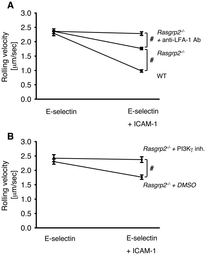
Rolling leukocytes are exposed to different adhesion molecules and chemokines. Neutrophils rolling on E-selectin induce integrin αLβ2-mediated slow rolling on ICAM-1 by activating a phospholipase C (PLC)γ2-dependent and a separate PI3Kγ-dependent pathway. E-selectin-signaling cooperates with chemokine signaling to recruit neutrophils into inflamed tissues. However, the distal signaling pathway linking PLCγ2 (Plcg2) to αLβ2-activation is unknown. To identify this pathway, we used different Tat-fusion-mutants and gene-deficient mice in intravital microscopy, autoperfused flow chamber, peritonitis, and biochemical studies. We found that the small GTPase Rap1 is activated following E-selectin engagement and that blocking Rap1a in Pik3cg-/- mice by a dominant-negative Tat-fusion mutant completely abolished E-selectin-mediated slow rolling. We identified CalDAG-GEFI (Rasgrp2) and p38 MAPK as key signaling intermediates between PLCγ2 and Rap1a. Gαi-independent leukocyte adhesion to and transmigration through endothelial cells in inflamed postcapillary venules of the cremaster muscle were completely abolished in Rasgrp2-/- mice. The physiological importance of CalDAG-GEFI in E-selectin-dependent integrin activation is shown by complete inhibition of neutrophil recruitment into the inflamed peritoneal cavity of Rasgrp2-/- leukocytes treated with pertussis toxin to block Gαi-signaling. Our data demonstrate that Rap1a activation by p38 MAPK and CalDAG-GEFI is involved in E-selectin-dependent slow rolling and leukocyte recruitment.


Conflict of interest statement

Conflict-of-interest disclosure

The authors declare no competing financial interests.

Figures

Figure 1
Figure 1. Rap1a is involved in E-selectin mediated slow rolling
(A) Bone-marrow-derived neutrophils from WT mice were lysed, treated with GTPγS (activator) or GDP (inactivator), and then purified using GST-RalGDS-RBD. Proteins were analyzed by Western blotting using a specific anti-Rap1 antibody. (B) Bone-marrow-derived neutrophils from WT mice were plated for different time intervals in multiwell plates with or without E-selectin coating, after which lysates were prepared. Western blots of activated Rap1 (affinity-precipitated Rap1-GTP) demonstrate Rap1 activation following E-selectin stimulation. Results are representative of 4 individual experiments. (C) Rap-1-GTP normalized to Rap1-GTP in unstimulated WT neutrophils (n=4). (D) Rolling velocities of reconstituted Lys-M-GFP+ leukocytes and Pik3cg−/− leukocytes pretreated with either wild type Rap1a TAT-peptides (Rap1-WT) or blocking Rap1a TAT-peptides (Rap1-DN) in inflamed postcapillary venules of the cremaster muscle of Lys-M-GFP+ mice and WT mice. The reconstituted cells were pretreated with PTx, and the mice were pretreated with anti–P-selectin mAb. Average rolling velocity of leukocytes presented as mean ± SEM. (D) Rolling velocities of reconstituted Plcg2−/− leukocytes pretreated with either wild type Rap1a TAT-peptides (Rap1-WT) or constitutively active Rap1a TAT-peptides (Rap1-CA) in inflamed postcapillary venules of the cremaster muscle of Lys-M-GFP+ mice. The reconstituted cells were pretreated with PTx, and the mice were pretreated with anti–P-selectin mAb. Average rolling velocity of leukocytes presented as mean ± SEM. #P < 0.05. * P < 0.05 vs. other groups.
Figure 2
Figure 2. Elimination of CalDAG-GEFI impairs E-selectin mediated slow rolling
(A) Carotid cannulas were placed in WT mice (n=3) and Rasgrp2−/− mice (n=3) and connected to autoperfused flow chambers. The wall shear stress in all flow chamber experiments was 5–6 dynes/cm2. (B) Rolling velocity of Rasgrp2−/− neutrophils on E-selectin alone or E-selectin/ICAM-1 of either PI3kγ-inhibitor (define molecule)- or DMSO-pretreated mice. Average rolling velocity of neutrophils on E-selectin (left) and E-selectin/ICAM-1 (right) presented as mean ± SEM. #P < 0.05.
Figure 3
Figure 3. CalDAG-GEFI and p38 MAPK are located in the PLCγ2-dependent signaling pathway
(A) Rolling velocity of WT neutrophils on E-selectin alone or E-selectin/ICAM-1 of either PLC inhibitor (U73122)- or DMSO-pretreated whole blood. (B) Rolling velocity of Rasgrp2−/− neutrophils on E-selectin alone or E-selectin plus ICAM-1 of either U73122- or DMSO-pretreated whole blood. (C) Rolling velocity of Rasgrp2−/− neutrophils on E-selectin alone or E-selectin/ICAM-1 of either p38 MAPK inhibitor (SB203580)– or DMSO-pretreated mice. Data presented as mean ± SEM from 3 mice. (D) (D) Whole human heparinized blood was treated with the p38 MAPK inhibitor SB203580 (10μM for 30 minutes at RT) or SB203580 (10μM) plus anti–LFA-1 antibody (10 μg/mL for 20 minutes at RT), then perfused through flow chambers coated with E-selectin with or without ICAM-1. Average rolling velocity of neutrophils on E-selectin (left) and E-selectin/ICAM-1 (right) is presented as mean ± SEM (n=3). (E) Whole human blood was treated with either Rap1-WT or Rap1-DN peptides (1μM for 30 minutes at RT), then perfused through flow chambers coated with E-selectin with or without ICAM-1. Average rolling velocity of neutrophils on E-selectin (left) and E-selectin/ICAM-1 (right) is presented as mean ± SEM (n=3). (F) HL-60 cells transfected with either siRNA specific for CalDAG-GEFI or a non-silencing control sequence were perfused through flow chambers coated with E-selectin and isotype antibody or KIM127 for 2 minutes at 5.94 dyn/cm2. The number of adherent cells per one representative field of view was determined. Data are from 3 experiments. #P < 0.05.
Figure 4
Figure 4. CalDAG-GEFI is involved in E-selectin mediated slow rolling in vivo
(A) Mixed chimeric mice were generated by injecting bone marrow cells from Rasgrp2−/− mice and LysM-GFP+ WT mice into lethally irradiated WT mice. Cumulative histogram of rolling velocity of 120 GFP+ leukocytes (wild-type, open circle), 120 GFP leukocytes (Rasgrp2−/−, filled triangle), and 120 Rasgrp2−/− leukocytes treated with a blocking LFA-1 antibody (Rasgrp2−/− + anti-LFA-1-antibody, open triangle) in inflamed cremaster muscle venules of mixed chimeric mice (n=4) treated with PTx and a monoclonal blocking P-selectin antibody. Insert: Mean ± SEM. #P<0.05. (B) Rolling velocities of reconstituted Lys-M-GFP+ leukocytes and Rasgrp2−/−- leukocytes pretreated with wild-type (WT) or constitutive-active (CA) Rap1a TAT-peptides in inflamed postcapillary venules of the cremaster muscle of Lys-M-GFP+ mice and WT mice. The reconstituted cells were pretreated with PTx, and the mice were pretreated with anti–P-selectin mAb. Average rolling velocity of leukocytes presented as mean ± SEM. * P < 0.05 vs. other groups. (C) Rap1a is involved in L-selectin clustering in rolling leukocytes. Wild-type leukocytes pretreated with either Rap1a-WT or Rap1a-DN peptides were injected in TNF-alpha treated mice before analysis of L-selectin redistribution by intravital microscopy. Results are derived from the analysis of three mice per group. #P<0.05.
Figure 5
Figure 5. PLCγ2 is upstream of CalDAG-GEFI and p38 MAPK and both are involved in Rap1 activation
Bone marrow–derived neutrophils from WT mice (untreated or pretreated with different inhibitors: phospholipase C: U73122, p38 MAPK: SB203580), Pik3cg−/− mice, Plcg2−/− mice, and Rasgrp2−/− mice were plated on uncoated (unstimulated) or E-selectin–coated wells, and then lysates were prepared. (A-C) Total Rap1 and GTP-bound Rap1 protein levels were measured in untreated or pretreated neutrophils from WT mice (A, untreated or pretreated with a phospholipase inhibitor (U73122)), Plcg2−/− mice (B), and Rasgrp2−/− mice (C) after stimulation with E-selectin. Representative blots from 3 independent experiments are shown. (D) Total Rap1 and GTP-bound Rap1 protein levels were measured in unstimulated and stimulated neutrophils from WT mice after inhibiting p38 MAPK (I, SB203580, 10μM). Representative blots from 3 independent experiments are shown. (E) Total Rap1 and GTP-bound Rap1 protein levels were measured in unstimulated and stimulated neutrophils from Pik3cg−/− mice. Representative blots from 3 independent experiments are shown. (F+G) Bone-marrow-derived neutrophils from WT mice (untreated or pretreated with a phospholipase inhibitor (U73122)) and Rasgrp2−/− mice were plated in multiwell plates with or without E-selectin coating for 10 minutes, after which lysates were prepared and immunoblotted with antibody to phosphorylated p38 MAPK (phospho-p38) or total p38. Representative blots from 3 independent experiments are shown.
Figure 6
Figure 6. Gαi-independent leukocyte adhesion, transmigration and recruitment is defective in Rasgrp2−/− mice
All mice were treated with pertussis toxin (PTx) to block Gαi-signaling. (A) Numbers of adherent cells per square millimeter in murine cremaster muscle venules. The cremaster muscle was exteriorized 2 hours after intrascrotal injection of 500 ng TNF-α in WT and Rasgrp2−/− mice. (B) Number of extravasated leukocytes in inflamed cremasteric venules of WT (n=3) and Rasgrp2−/− mice (n=3) per 1.5 × 104 μm2 tissue area. The measurements were performed 2 hours after intrascrotal TNF-α injection. (C+D) Representative reflected light oblique transillumination microscopic pictures of cremaster muscle postcapillary venules of PTx pretreated WT mice (C) and Rasgrp2−/− mice (D) 2 h after TNF-α application. Demarcations on each side of the venule determine the areas in which extravasated leukocytes were counted. Scale bar equals 50 μm. (E) Neutrophil influx into the peritoneal cavity 8 hrs after 1 ml injection of 4% thioglycollate into mixed chimeric mice generated by injecting bone marrow from WT mice and Rasgrp2−/− mice into lethally irradiated WT mice. After 6 weeks, , mice received 4μg PTx i.v. to block Gαi-signaling followed bye thioglycollate injection. Migration efficiency was calculated (number of recruited neutrophils/number of neutrophils in the blood). Data presented as mean ± SEM from 5 mice. #P < 0.05.

Similar articles

Cited by

References

    1. Anderson DC, Springer TA. Leukocyte adhesion deficiency: an inherited defect in the Mac-1, LFA-1, and p150,95 glycoproteins. Annu Rev Med. 1987;38:175–194. - PubMed
    1. Nathan C. Neutrophils and immunity: challenges and opportunities. Nat Rev Immunol. 2006;6:173–182. - PubMed
    1. Butcher EC. Leukocyte-endothelial cell recognition: three (or more) steps to specificity and diversity. Cell. 1991;67:1033–1036. - PubMed
    1. Springer TA. Traffic signals for lymphocyte recirculation and leukocyte emigration: the multistep paradigm. Cell. 1994;76:301–314. - PubMed
    1. Ley K, Laudanna C, Cybulsky MI, Nourshargh S. Getting to the site of inflammation: the leukocyte adhesion cascade updated. Nat Rev Immunol. 2007;7:678–689. - PubMed

Publication types

MeSH terms

Substances

LinkOut - more resources