. 2011 Apr;53(4):1323-32.
doi: 10.1002/hep.24203.

Fibrinogen-γ proteolysis and solubility dynamics during apoptotic mouse liver injury: heparin prevents and treats liver damage

Affiliations

Fibrinogen-γ proteolysis and solubility dynamics during apoptotic mouse liver injury: heparin prevents and treats liver damage

Sujith V W Weerasinghe et al. Hepatology. 2011 Apr.

Abstract

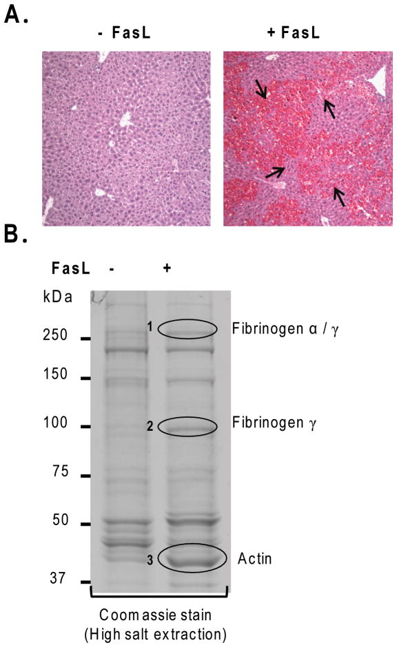
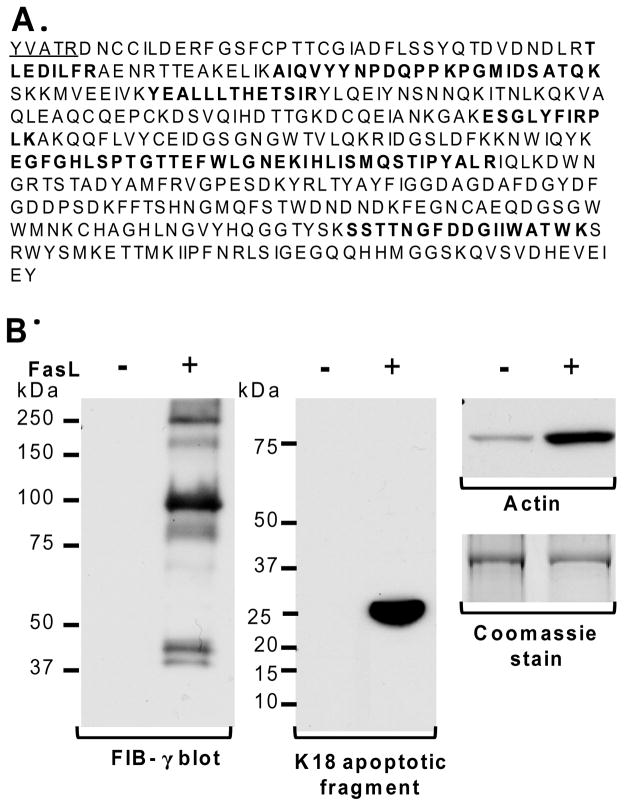
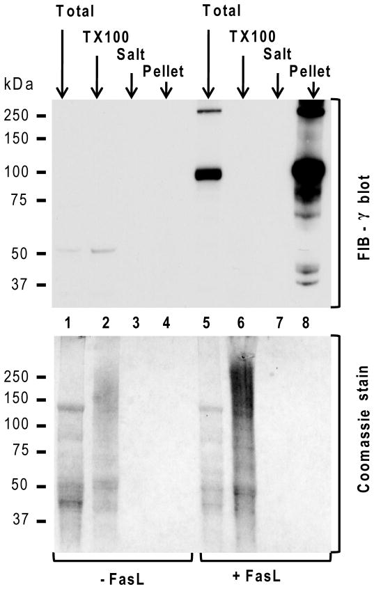
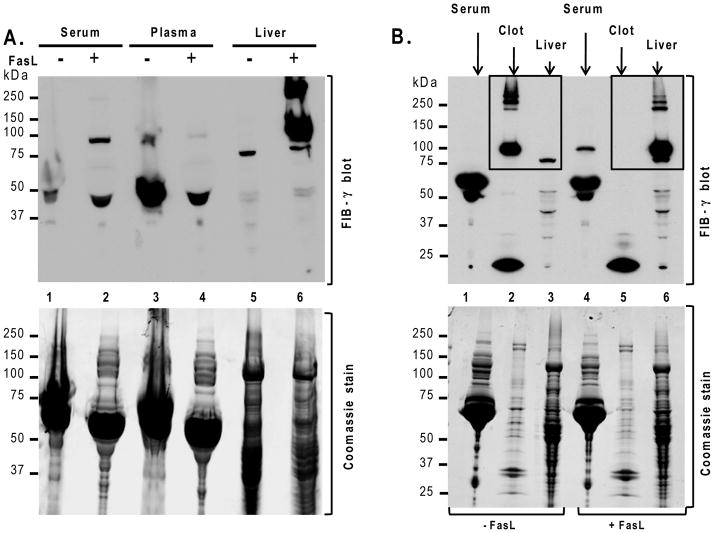
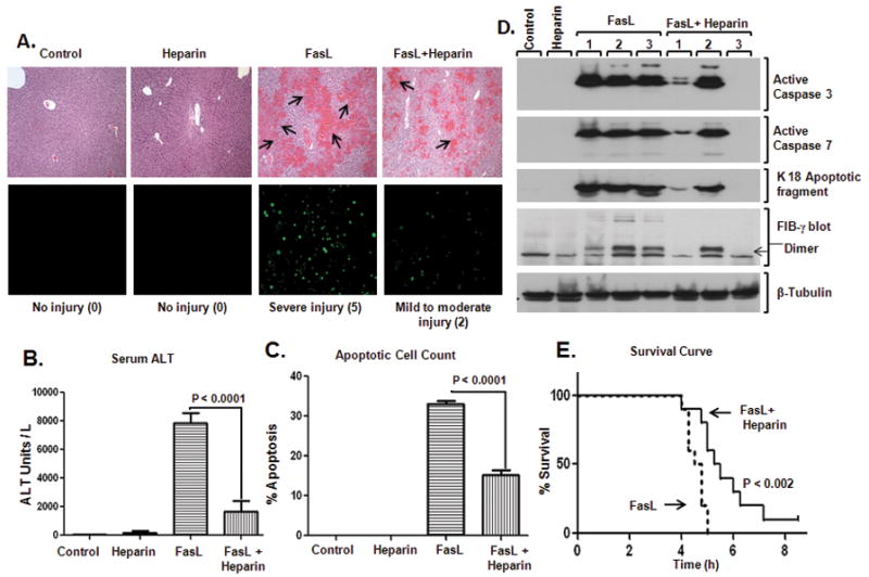
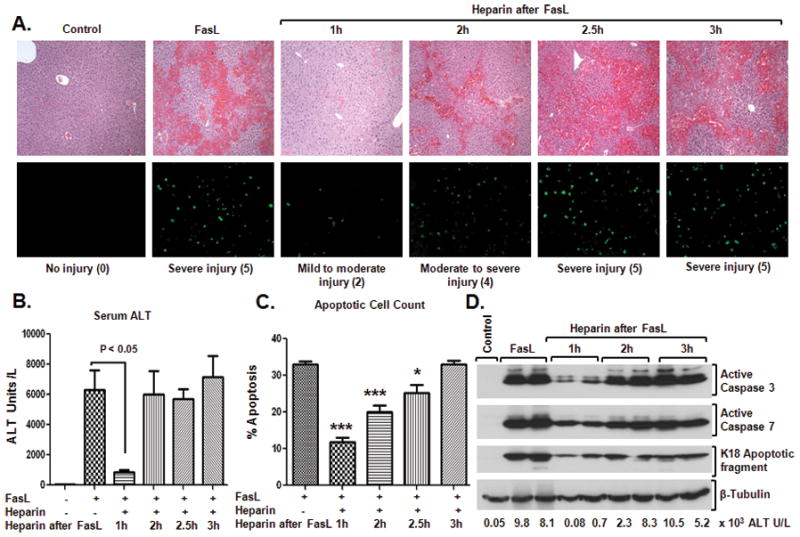
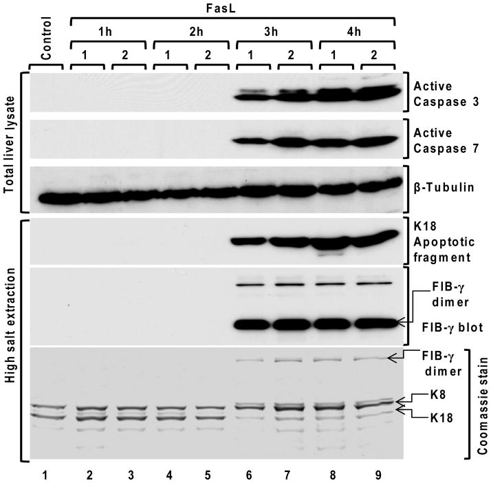
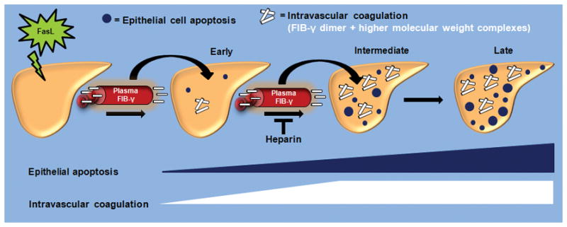
Fas ligand (FasL)-mediated hepatocyte apoptosis occurs in the context of acute liver injury that can be accompanied by intravascular coagulation (IC). We tested the hypothesis that analysis of selected protein fractions from livers undergoing apoptosis will shed light on mechanisms that are involved in liver injury that might be amenable to intervention. Proteomic analysis of the major insoluble liver proteins after FasL exposure for 4-5 hours identified fibrinogen-γ (FIB-γ) dimers and FIB-γ-containing high molecular mass complexes among the major insoluble proteins visible via Coomassie blue staining. Presence of the FIB-γ-containing products was confirmed using FIB-γ-specific antibodies. The FIB-γ-containing products partition selectively and quantitatively into the liver parenchyma after inducing apoptosis. Similar formation of FIB-γ products occurs after acetaminophen administration. The observed intrahepatic IC raised the possibility that heparin therapy may ameliorate FasL-mediated liver injury. Notably, heparin administration in mice 4 hours before or up to 2 hours after FasL injection resulted in a dramatic reduction of liver injury-including liver hemorrhage, serum alanine aminotransferase, caspase activation, and liver apoptosis-compared with heparin-untreated mice. Heparin did not directly interfere with FasL-induced apoptosis in isolated hepatocytes, and heparin-treated mice survived the FasL-induced liver injury longer compared with heparin-untreated animals. There was a sharp, near-simultaneous rise in FasL-induced intrahepatic apoptosis and coagulation, with IC remaining stable while apoptosis continued to increase.

Conclusion: Formation of FIB-γ dimers and their high molecular mass products are readily detectable within the liver during mouse apoptotic liver injury. Heparin provides a potential therapeutic modality, because it not only prevents extensive FasL-related liver injury but also limits the extent of injury if given at early stages of injury exposure.


Figures

Figure 1
Figure 1. Characterization of the insoluble liver proteins after FasL-induced apoptosis
(A) FasL was administered to mice. After 4.5h, mice were sacrificed followed by H&E staining. Note the dramatic hemorrhage (arrows) in FasL-treated compared to untreated mice. (B) Insoluble protein fractions of livers from FasL-treated and untreated mice were isolated using HSE followed by SDS-PAGE and Coomassie staining. Bands that showed increased intensity after FasL-treatment (circles 1,2,3) were subjected to mass spectrometric analysis which, in turn, predicted the identity of bands 1–3 as: fibrinogen α/γ(1), fibrinogen-γ(2) and actin (3).
Figure 2
Figure 2. FIB-γ is the major insoluble liver protein after FasL-mediated injury
(A) Fragment sequences of FIB-γ obtained from N-terminal sequencing (underlined) and mass spectrometry (bold) confirm the identity of band #2 shown in Fig. 1B. (B) Prediction of mass spectrometric results was verified by immunoblotting using antibodies to FIB-γ and actin. The FIB-γ antibody recognized several bands in the FasL-treated liver (250-kDa and 100-kDa) that were not present in the untreated controls. These species correspond to bands 1 and 2 (Fig. 1B), respectively. The actin blot demonstrated elevated levels of actin protein in FasL-treated liver compared to untreated control. Formation of the K18 apoptotic fragment confirmed the hepatocyte FasL-induced apoptosis.
Figure 3
Figure 3. Solubility dynamics of FIB-γ during FasL-induced liver injury
Solubility dynamics of FIB-γ during FasL-induced liver injury were analyzed biochemically by comparing the soluble TX100 fraction, high salt wash fraction (Salt) and the insoluble fraction (Pellet) from FasL-treated and untreated livers. TLL (Total) were used as controls. None of the fractions showed detectable levels of FIB-γ in untreated liver samples. However, FIB-γ antibody faintly recognized the FIB-γ monomer (50-kDa) in the TLL and the soluble TX100 fraction of untreated liver. In contrast, FIB-γ blot showed two major bands (100-kDa, 250-kDa) in FasL-treated TLL, but not in the soluble TX100 and wash fractions. A duplicate gel to that used for immunoblotting was stained with Coomassie blue. Athough the protein amounts in some of the lanes (3,4,7,8) were not enough to be visible with Coomassie staining, they were readily detected by the FIB-γ antibody (lane 8).
Figure 4
Figure 4. Intrahepatic accumulation of FIB-γ during FasL-induced injury
Mice were sacrificed 4.5h after FasL-injection and blood components (serum, plasma, clot) and liver were collected. (A) FIB-γ antibody recognized the FIB-γ dimer (100-kDa) in FasL-treated mouse serum but not in FasL-untreated serum. FIB-γ dimers and other HMW products (~250-kDa) were readily observed in FasL-treated liver lysates. (B) FIB-γ dimers and other HMW products were clearly observed by immunoblotting in the clot fraction of untreated mice but not in the liver, while the opposite occurs after FasL treatment. Coomassie staining of duplicate gels to those used for blotting is included as loading control.
Figure 5
Figure 5. Pretreatment with heparin reduces FasL-induced liver injury
(A) Heparin was administered to age/sex-matched mice followed by FasL injection (4h after heparin), and the extent of liver damage was assessed. H&E sections show hemorrhage formation (top) and TUNEL staining shows apoptotic cells (bottom). Three mice were used for each of the control and heparin-alone arms, while 7 mice were used for each of the FasL and FasL+heparin arms. Numbers in parenthesis reflect the injury score (see Experimental Procedures and Fig. S2). (B) Serum ALT, (C) % Apoptotic cell count, and (D) Biochemical analysis of the livers from the indicated treatments and controls are shown. Note that all measured parameters are significantly less in mice that received heparin prior to FasL. In panel D, each lane represents an independent liver, and tubulin is shown as a loading control. (E) Heparin or saline were administered to mice (10–12 week-old; n=10/group) followed by FasL-injection (4h after heparin/saline) and survival time (hours) was recorded.
Figure 6
Figure 6. Early treatment with heparin reduces FasL-induced liver injury
FasL was administered intraperitoneally followed by heparin injection 1h, 2h, 2.5h and 3h after FasL (5–7 mice/time point). After 4.5h of FasL injection, mice were sacrificed and liver damage was assessed. (A) H&E staining (top) and TUNEL staining (bottom) showed that treatment with heparin at 1h and 2h after FasL injection significantly reduced hemorrhage formation and apoptosis, indicative of only mild-to-moderate injury, as compared to heparin-untreated mice. Numbers in parenthesis reflect the injury score (see Experimental Procedures and Fig. S5). (B) and (C) show serum ALT levels and percentage of apoptotic cells, respectively. (D) Activation of caspase-3 and 7 is markedly reduced in mice that received heparin 1h following FasL-injection. Consistent with the decreased caspase activation, lower levels of the K18 apoptotic fragment were detected in livers of mice that received heparin treatment 1h following FasL injection as compared with mice that received FasL-alone. Serum ALT levels for each of the biochemically analyzed livers of each animal are shown. Tubulin blot is included as a loading control.
Figure 7
Figure 7. Time course of caspase activation, K18 caspase-mediated digestion and FIB-γ dimer formation after FasL-induced injury
Liver apoptosis was induced by FasL-injection. Mice were sacrificed at 1h, 2h, 3h and 4h after FasL-injection (2 mice/time point), followed by preparation of the total liver lysates and HSE then immunoblotting using antibodies to the indicated antigens. A tubulin blot is included as a loading control, and a Coomassie stain of a duplicate gel of the analyzed HSE samples that were analyzed by blotting is also included.
Figure 8
Figure 8. Model of intrahepatic vascular coagulation and epithelial cell apoptosis dynamics in response to FasL
The early stages of FasL-induced liver injury are manifested by limited hepatocytes apoptosis and intrahepatic vascular coagulation. This is followed at the intermediate stages by a near-concurrent marked increase in liver caspase activation, epithelial apoptosis and intravascular coagulation. The IC is manifested by the formation of FIB-γ dimers and HMW products within the liver, in association with a dramatic shift of FIB-γ from plasma to liver. During the late stages that follow FasL exposure, IC remains relatively unchanged while hepatocytes apoptosis continues. Heparin provides a therapeutic modality that not only limits the extensive of FasL-related liver injury when given as prophylactic but also limits the extent of injury if given at the early stages of injury exposure.

References

    1. Malhi H, Gores GJ, Lemasters JJ. Apoptosis and necrosis in the liver: a tale of two deaths? Hepatology. 2006;43:S31–44. - PubMed
    1. Malhi H, Guicciardi ME, Gores JG. Hepatocyte Death: A Clear and Present Danger. Physiol Rev. 2010;90:1165–1194. - PMC - PubMed
    1. Ku NO, Liao J, Omary MB. Apoptosis generates stable fragments of human type I keratins. J Biol Chem. 1997;272:33197–33203. - PubMed
    1. Caulin C, Salvesen GS, Oshima RG. Caspase cleavage of keratin 18 and reorganization of intermediate filaments during epithelial cell apoptosis. J Cell Biol. 1997;138:1379–1394. - PMC - PubMed
    1. Rao L, Perez D, White E. Lamin proteolysis facilitates nuclear events during apoptosis. J Cell Biol. 1996;135:1441–1455. - PMC - PubMed

Publication types