. 2011 Jun;32(6):669-77.
doi: 10.1002/humu.21488. Epub 2011 Apr 7.

A complex phenotype of peripheral neuropathy, myopathy, hoarseness, and hearing loss is linked to an autosomal dominant mutation in MYH14

Affiliations

A complex phenotype of peripheral neuropathy, myopathy, hoarseness, and hearing loss is linked to an autosomal dominant mutation in MYH14

Byung-Ok Choi et al. Hum Mutat. 2011 Jun.

Abstract

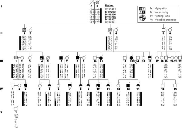
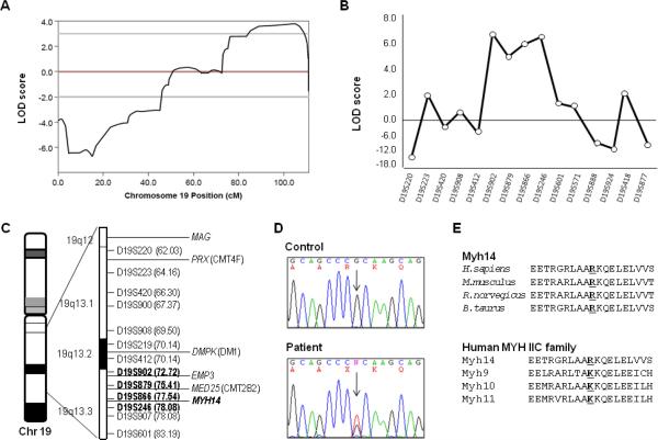
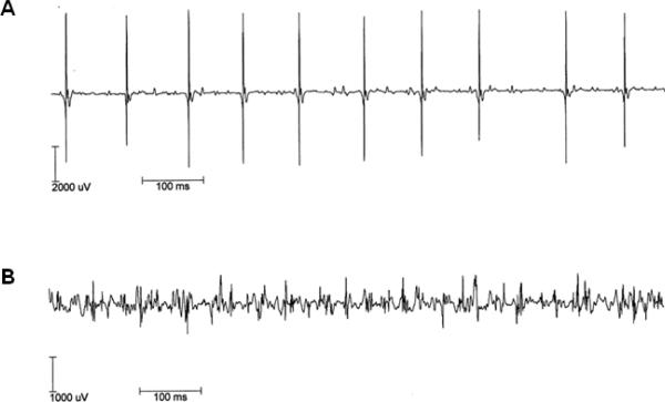
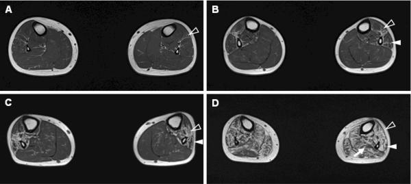
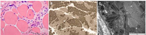
Both peripheral neuropathy and distal myopathy are well-established inherited neuromuscular disorders characterized by progressive weakness and atrophy of the distal limb muscles. A complex phenotype of peripheral neuropathy, myopathy, hoarseness, and hearing loss was diagnosed in a large autosomal dominant Korean family. A high density single nucleotide polymorphism (SNP)-based linkage study mapped the underlying gene to a region on chromosome 19q13.3. The maximum multipoint LOD score was 3.794. Sequencing of 34 positional candidate genes in the segregating haplotype revealed a novel c.2822G>T (p.Arg941Leu) mutation in the gene MYH14, which encodes the nonmuscle myosin heavy chain 14. Clinically we observed a sequential pattern of the onset of muscle weakness starting from the anterior to the posterior leg muscle compartments followed by involvement of intrinsic hand and proximal muscles. The hearing loss and hoarseness followed the onset of distal muscle weakness. Histopathologic and electrodiagnostic studies revealed both chronic neuropathic and myopathic features in the affected patients. Although mutations in MYH14 have been shown to cause nonsyndromic autosomal dominant hearing loss (DFNA4), the peripheral neuropathy, myopathy, and hoarseness have not been associated with MYH14. Therefore, we suggest that the identified mutation in MYH14 significantly expands the phenotypic spectrum of this gene.


Figures

Figure 1
Figure 1
Pedigree of family FC317. Haplotypes (indicated below each familial member) were determined using genotypes of microsatellite markers at chromosome 19q13.3 (genotypes in parentheses were inferred). Asterisks (*) indicate individuals whose DNA was used for linkage analysis. Black color indicates confirmed phenotypes by clinical exam, and grey indicates presumptive phenotypes based on history talking. Arrow (↖) indicates the proband.
Figure 2
Figure 2
Linkage analysis of family FC317 and identification of a missense mutation in MYH14 gene. (A) Two-point LOD scores of chromosome 19. (B) Chromosomal fine mapping of the 19q13.2-3 region. Genotyping of 29 microsatellites revealed a ~13Mb linkage region flanked by markers D19S902 and D19S246. (C) Diagram of chromosome 19q13.2-3. Markers that co-segregate with the phenotype are indicated in bold. (D) Sequencing chromatograms of the c.2822G>T (Arg941Leu) mutation in the MYH14 gene. The variant co-segregated with the affected family members. (E) High conservation of the Arg94 residue is illustrated by alignment of the amino acid sequences of MYH14 from different species and the MYH IIC family of proteins (ClustalX program, ver. 1.83) (H. sapiens MYH14-NP_001070654.1, MYH9-NP_002464.1, MYH10-NP_005955.1, MYH11-NP_002465.1; M. musculus Myh14-NP_082297.1, R. norvegicus Myh14- NP_001094160.1, B. Taurus Myh14- XM_882711.4).
Figure 3
Figure 3
Motor unit action potential (MUAP). Recordings of the interference patterns in patient III-8 were made from the right vastus lateralis (A), and the first dorsal interosseous (B). The patient has evidence, clinically and electrophysiologically, of both neuropathy and myopathy. His electrophisiological studies revealed both a large amplitude with long duration (neuropathic, A) and a small amplitude with short duration polyphasic (myopathic) motor units (B).
Figure 4
Figure 4
A sequential pattern of muscle involvement associated with disease duration (DD) in the T1-weighted axial MRI. (A) A 16-yr-old male patient (IV-10, DD=4 yrs) showed mild streaky fatty infiltrations of the anterior compartment muscles of the legs. (B) A 15-yr-old male patient (IV-13, DD=5 yrs) displayed more fatty infiltrations, including the anterior and the lateral compartments. (C) A 33-yr-old male (III-16, DD=24 yrs) showed prominent involvement of the anterior and lateral compartments of leg muscles with mild involvement of the posterior compartment. (D) A 41-yr-old female patient (III-13, DD=28 yrs) showed severe fatty involvement of all muscles compartments including a calf muscle atrophy (anterior compartment: blank arrowheads, lateral compartment: arrowheads, posterior compartment: arrow).
Figure 5
Figure 5
Histopathologic findings of lateral gastrocnemius muscle in patient III-16. (A) A frozen section showing marked variation of fiber size and shape with many small rounded fibers, and degenerating fibers. Endomysial fibrosis was prominent on the background of degenerating myofibers without inflammatory cell infiltration by H&E stain. (B) Grouping of histochemical fiber types. ATPase reaction with different pH preincubation and immunostaining with myosin heavy chain (fast), myosin heavy chain (slow), and myosin IIa showed grouping of histochemical fiber types. (C) Subsarcolemmal accumulation of abnormal mitochondria. Electron micrographs revealed subsarcolemmal accumulation of frequent abnormal mitochondria including variable sized rectangular or elongated rhomboidal paracrystalline inclusions. Magnification: × 200 (A), × 40 (B), and × 30,000 (C).

References

    1. Arias M, Pardo J, Blanco-Arias P, Sobrido MJ, Arias S, Dapena D, Carracedo A, Goldfarb LG, Navarro C. Distinct phenotypic features and gender-specific disease manifestations in a Spanish family with desmin L370P mutation. Neuromuscul Disord. 2006;16:498–503. - PubMed
    1. Astuto LM, Kelley PM, Askew JW, Weston MD, Smith RJ, Alswaid AF, Al-Rakaf M, Kimberling WJ. Searching for evidence of DFNB2. Am J Med Genet. 2002;109:291–297. - PubMed
    1. Berghoff C, Berghoff M, Leal A, Morera B, Barrantes R, Reis A, Neundörfer B, Rautenstrauss B, Del Valle G, Heuss D. Clinical and electrophysiological characteristics of autosomal recessive axonal Charcot-Marie-Tooth disease (ARCMT2B) that maps to chromosome 19q13.3. Neuromuscul Disord. 2004;5:301–306. - PubMed
    1. Bergmann C, Senderek J, Hermanns B, Jauch A, Janssen B, Schröder JM, Karch D. Becker muscular dystrophy combined with X-linked Charcot-Marie-Tooth neuropathy. Muscle Nerve. 2000;23:818–823. - PubMed
    1. Braida C, Stefanatos RK, Adam B, Mahajan N, Smeets HJ, Niel F, Goizet C, Arveiler B, Koenig M, Lagier-Tourenne C, et al. Variant CCG and GGC repeats within the CTG expansion dramatically modify mutational dynamics and likely contribute toward unusual symptoms in some myotonic dystrophy type 1 patients. Hum Mol Genet. 2010;19:1399–1412. - PubMed

Publication types

MeSH terms