Review
. 2011 Jun;125(3):282-96.
doi: 10.1037/a0023165.

Molecular modulation of prefrontal cortex: rational development of treatments for psychiatric disorders

Affiliations
Review

Molecular modulation of prefrontal cortex: rational development of treatments for psychiatric disorders

Nao J Gamo et al. Behav Neurosci. 2011 Jun.

Abstract

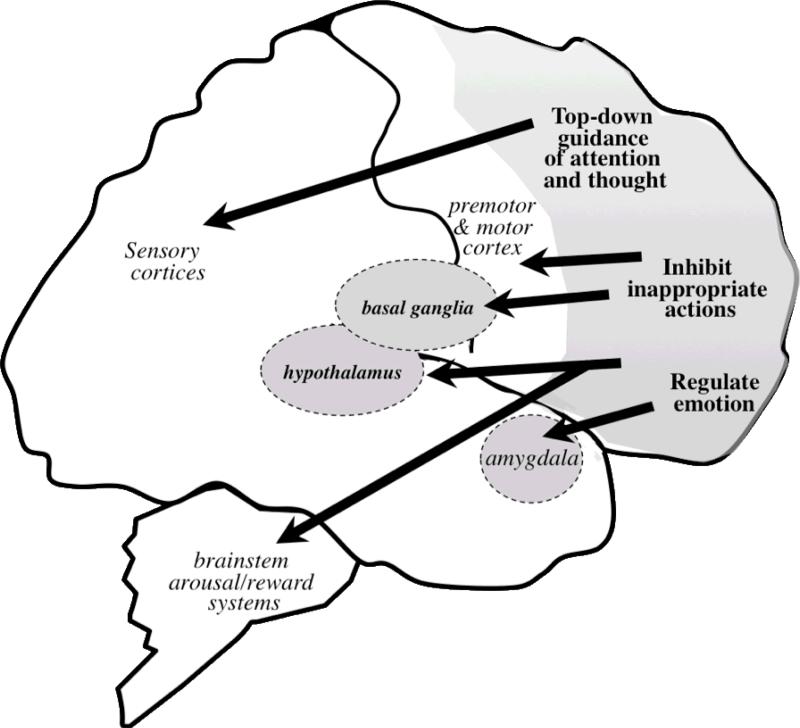
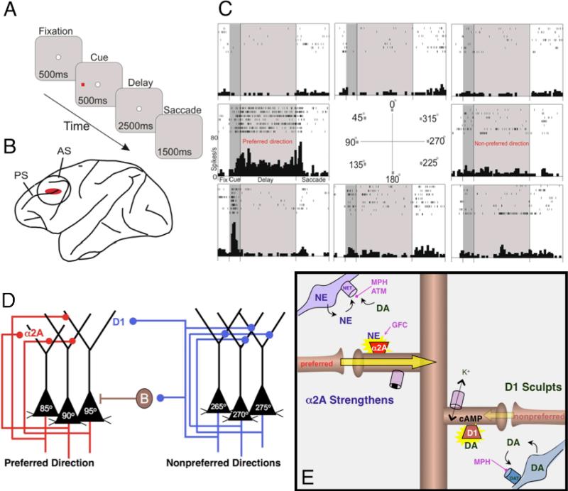
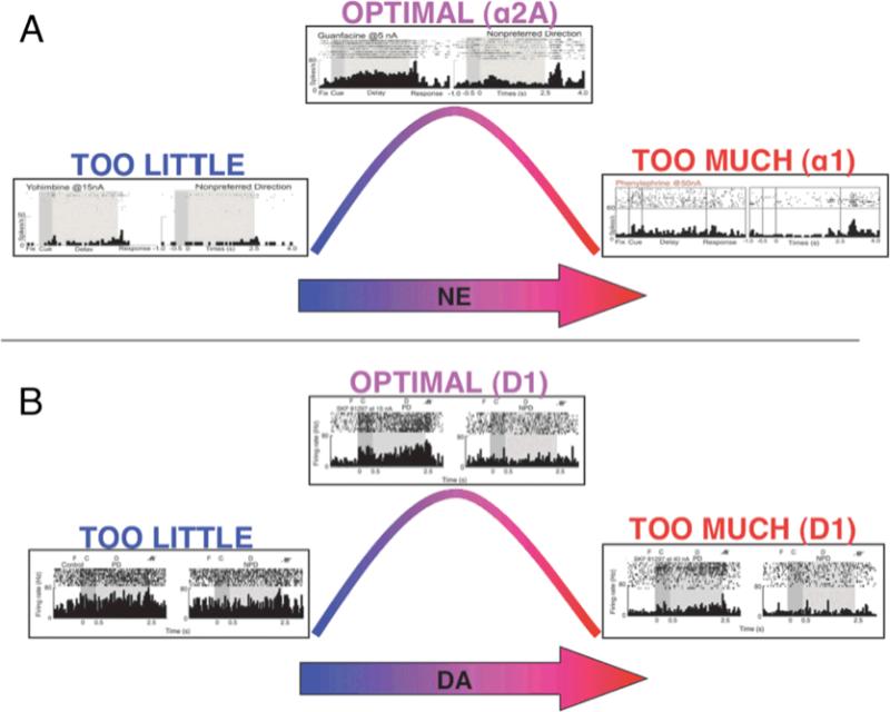
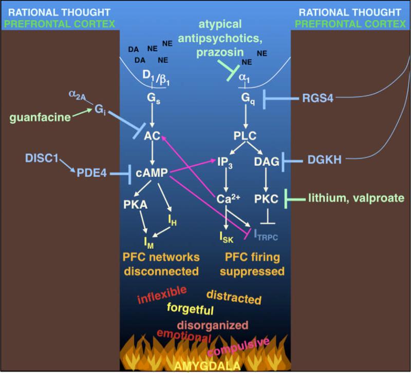
Dysfunction of the prefrontal cortex (PFC) is a central feature of many psychiatric disorders, such as attention deficit hyperactivity disorder (ADHD), posttraumatic stress disorder (PTSD), schizophrenia, and bipolar disorder. Thus, understanding molecular influences on PFC function through basic research in animals is essential to rational drug development. In this review, we discuss the molecular signaling events initiated by norepinephrine and dopamine that strengthen working memory function mediated by the dorsolateral PFC under optimal conditions, and weaken working memory function during uncontrollable stress. We also discuss how these intracellular mechanisms can be compromised in psychiatric disorders, and how novel treatments based on these findings may restore a molecular environment conducive to PFC regulation of behavior, thought and emotion. Examples of successful translation from animals to humans include guanfacine for the treatment of ADHD and related PFC disorders, and prazosin for the treatment of PTSD.


Figures

Figure 1
Figure 1
The prefrontal cortex (PFC) is highly interconnected with the rest of the brain, allowing it to modulate information processing in other regions of the brain, as well as to regulate the arousal and reward systems, to appropriately modulate behavior, thought and affect.
Figure 2
Figure 2
Physiological recordings of PFC microcircuits in the monkey dorsolateral PFC (DLPFC), and their modulation by catecholamine signaling pathways. A. Monkeys performed the oculomotor delayed response (ODR) task. Each trial begins when the monkey fixates on a central point on a screen (fixation period). Next, a cue briefly appears in 1 of 8 peripheral locations, followed by a 2.5-second delay period, during which the monkey continues to maintain fixation. At the end of the delay period, the monkey makes a memory-guided saccade to the remembered cue location (response period), and is rewarded with juice if correct. Each test session consists of hundreds of trials, in which the cued location randomly changes for each trial, thus requiring the monkey to update his working memory despite extensive, proactive interference. B. Single-unit recording was performed in the DLPFC, the region associated with spatial working memory in monkeys. C. The firing patterns of a representative neuron in the monkey DLPFC. Under optimal conditions, neurons show delay-related firing for a preferred direction, but suppress firing for non-preferred directions (Goldman-Rakic, 1995; Goldman-Rakic, 1996). This pattern is considered to be the cellular basis for spatial working memory. D. Circuit basis for spatial working memory as proposed by Patricia Goldman-Rakic (Goldman-Rakic, 1995). Spatial working memory is maintained in the DLPFC by recurrent excitation among networks of glutamatergic pyramidal cells with shared stimulus inputs, e.g. 90° position in space. Spatial tuning is sharpened by GABAergic interneurons, such as basket cells (B) (Rao et al., 1999; Rao et al., 2000), that suppress firing for dissimilar spatial positions, e.g. 270°. The preferred inputs to the 90° neuron are shown in red; physiological data indicate that these networks are modulated by norepinephrine (NE) alpha-2A adrenoceptor stimulation. In contrast, inputs from non-preferred directions are shown in blue; physiological data indicate that these networks are sculpted by D1 dopamine (DA) receptor stimulation. E. A working model of catecholamine influences on PFC network connectivity. Under optimal levels of PFC catecholamine release, alpha-2A adrenoceptors strengthen connectivity for the preferred direction of the PFC network by reducing cyclic adenosine monophosphate (cAMP) and closing hyperpolarizing potassium channels. Conversely, D1 DA receptors sharpen spatial tuning by weakening inputs from non-preferred directions by increasing cAMP production. Treatments for ADHD facilitate these actions to restore optimal PFC function, e.g. methylphenidate (MPH) and atomoxetine (ATM) block NE and DA transporters (NET and DAT) and enhance the endogenous stimulation of alpha-2A and D1 receptors, while guanfacine (GFC) directly stimulates alpha-2A adrenoceptors.
Figure 3
Figure 3
NE and DA show inverted-U dose-response curves on delay-related activity in a monkey performing the ODR task. A. Optimal levels of NE enhance delay-related activity for the preferred direction via alpha-2A adrenoceptors, while excessive levels suppress firing via alpha-1 and beta-1 adrenoceptors (Birnbaum et al., 2004; Wang et al., 2007). B. Optimal levels of DA enhance spatial tuning by suppressing delay-related firing for the non-preferred direction, while excessive levels suppress firing for both preferred and non-preferred stimuli (Vijayraghavan et al., 2007).
Figure 4
Figure 4
During exposure to uncontrollable stress, high levels of NE and DA release initiate intracellular signaling events that increase potassium currents (IH, IM and ISK), and decrease depolarizing currents (ICAN, also called ITRPC). This cascade leads to a collapse in PFC network firing and a loss of PFC regulation of behavior. Many of these signaling events appear to have feed-forward interactions, e.g. whereby cAMP facilitates IP3 receptor signaling and vice versa, thus providing the opportunity for rapid changes in cellular physiology. In such a situation, rational control of behavior goes to “hell in a handbasket,” and is replaced by instinctual reactions mediated by subcortical regions such as the amygdala, which are strengthened rather than weakened by PKA and PKC signaling. However, there are signaling pathways in the PFC that serve as “molecular brakes” to inhibit these stress-induced pathways, e.g. DISC1, RGS4 and DGK. Intriguingly, these molecules are often genetically altered in mental illness. Many treatments for psychiatric disorders inhibit these stress responses, e.g. prazosin and atypical antipsychotics block alpha-1 adrenoceptors, while the bipolar agents, lithium and valproate, reduce PKC signaling. These treatments may restore a more optimal signaling environment in the PFC. Abbreviations: AC: adenylyl cyclase; cAMP: cyclic adenosine monophosphate; DA: dopamine; DAG: diacylglycerol; DGKH: DAG kinase-η; DISC1: Disrupted In Schizophrenia 1; IH: h-current mediated by hyperpolarization-activated cyclic nucleotide gated cation (HCN) channels; IM: M-current mediated by KCNQ channels; IP3: inositol triphosphate; ISK: current mediated by small-conductance calcium-activated potassium (SK) channels; ITRPC: current mediated by canonical transient receptor potential channels (TRPC); NE: norepinephrine; PDE4: phosphodiesterase 4; PFC: prefrontal cortex; PKA: protein kinase A; PKC: protein kinase C; PLC: phopholipase C; RGS4: Regulator of G-protein Signaling 4.

References

    1. Aoki C, Go CG, Venkatesan C, Kurose H. Perikaryal and synaptic localization of alpha 2A-adrenergic receptor-like immunoreactivity. Brain Research. 1994;650(2):181–204. - PubMed
    1. Aoki C, Venkatesan C, Kurose H. Noradrenergic modulation of the prefrontal cortex as revealed by electron microscopic immunocytochemistry. Advances in Pharmacology (San Diego, Calif.) 1998;42:777–780. - PubMed
    1. Arnsten AFT, Castellanos FX. Neurobiology of attention regulation and its disorders. In: Martin A, Scahill L, Charney D, Leckman J, editors. Textbook of child and adolescent pharmacology. Oxford University Press; New York, NY: 2002. pp. 99–109.
    1. Arnsten AFT, Manji HK. Mania: A rational neurobiology. Future Neurology. 2008;3(2):125–131.
    1. Arnsten AFT, Vijayraghavan S, Wang M, Gamo NJ, Paspalas CD. Dopamine's influence on prefrontal cortical cognition: Actions and circuits in behaving primates. In: Iversen L, Iversen S, Dunnett S, Bjorklund A, editors. Dopamine handbook. 1st ed. Oxford University Press; New York, NY: 2009. pp. 230–248.

MeSH terms