

An ear punch model for studying the effect of radiation on wound healing
- PMID: 21480768
- PMCID: PMC3193941
- DOI: 10.3109/09553002.2011.568575
An ear punch model for studying the effect of radiation on wound healing
Abstract
Purpose: Radiation and wound combined injury represents a major clinical challenge because of the synergistic interactions that lead to higher morbidity and mortality than either insult would produce singly. The purpose of this study was to develop a mouse ear punch model to study the physiological mechanisms underlying radiation effects on healing wounds.
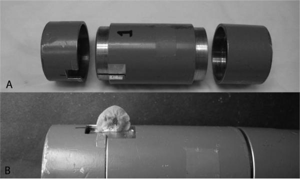
Materials and methods: Surgical wounds were induced by a 2 mm surgical punch in the ear pinnae of MRL/MpJ mice. Photographs of the wounds were taken and the sizes of the ear punch wounds were quantified by image analysis. Local radiation to the ear was delivered by orthovoltage X-ray irradiator using a specially constructed jig that shields the other parts of body.
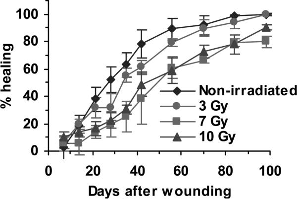
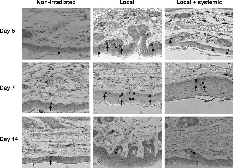
Results: Using this model, we demonstrated that local radiation to the wound area significantly delayed the healing of ear punch wounds in a dose-dependent fashion. The addition of sublethal whole body irradiation (7 Gy) further delayed the healing of ear punch wounds. These results were replicated in C57BL/6 mice; however, wound healing in MRL/MpJ mice was accelerated.
Conclusions: These data indicate that the mouse ear punch model is a valuable model to study radiation and wound combined injury.
Figures


Similar articles
-
Regeneration of the ear after wounding in different mouse strains is dependent on the severity of wound trauma.Dev Dyn. 2003 Feb;226(2):388-97. doi: 10.1002/dvdy.10242. Dev Dyn. 2003. PMID: 12557217
-
Denervation affects regenerative responses in MRL/MpJ and repair in C57BL/6 ear wounds.J Anat. 2012 Jan;220(1):3-12. doi: 10.1111/j.1469-7580.2011.01452.x. Epub 2011 Nov 8. J Anat. 2012. PMID: 22066944 Free PMC article.
-
Peripheral nerve regeneration in the MRL/MpJ ear wound model.J Anat. 2011 Feb;218(2):163-72. doi: 10.1111/j.1469-7580.2010.01313.x. Epub 2010 Oct 18. J Anat. 2011. PMID: 20950365 Free PMC article.
-
Characterizing regeneration in the vertebrate ear.J Anat. 2006 Oct;209(4):439-46. doi: 10.1111/j.1469-7580.2006.00632.x. J Anat. 2006. PMID: 17005017 Free PMC article. Review.
-
Regeneration of articular cartilage in healer and non-healer mice.Matrix Biol. 2014 Oct;39:50-5. doi: 10.1016/j.matbio.2014.08.011. Epub 2014 Aug 28. Matrix Biol. 2014. PMID: 25173437 Free PMC article. Review.
Cited by
-
Short-term influences of radiation on musculofascial healing in a laparotomy rat model.Sci Rep. 2019 Aug 15;9(1):11896. doi: 10.1038/s41598-019-48201-5. Sci Rep. 2019. PMID: 31417127 Free PMC article.
-
Inhibition of apoptosis signal-regulating kinase 1 alters the wound epidermis and enhances auricular cartilage regeneration.PLoS One. 2017 Oct 18;12(10):e0185803. doi: 10.1371/journal.pone.0185803. eCollection 2017. PLoS One. 2017. PMID: 29045420 Free PMC article.
-
Increase in the radioresistance of normal skin fibroblasts but not tumor cells by mechanical injury.Cell Death Dis. 2017 Feb 2;8(2):e2573. doi: 10.1038/cddis.2016.416. Cell Death Dis. 2017. PMID: 28151479 Free PMC article.
References
-
- Baxter H, Drummond JA, Stephens-Newsham LG, Randall RG. Studies on acute total body irradiation in animals. I. Effect of streptomycin following exposure to a thermal burn and irradiation. Plastic Reconstructive Surgery. 1953;12:439–445. 1946. - PubMed
Publication types
MeSH terms
Grants and funding
LinkOut - more resources
Full Text Sources