. 2011 Apr 12:8:163.
doi: 10.1186/1743-422X-8-163.

Lysosomotropic agents as HCV entry inhibitors

Affiliations

Lysosomotropic agents as HCV entry inhibitors

Usman A Ashfaq et al. Virol J. .

Abstract

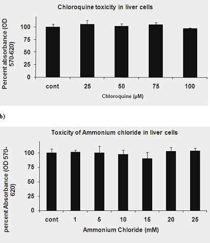
HCV has two envelop proteins named as E1 and E2 which play an important role in cell entry through two main pathways: direct fusion at the plasma membrane and receptor-mediated endocytosis. Fusion of the HCV envelope proteins is triggered by low pH within the endosome. Lysosomotropic agents (LA) such as Chloroquine and Ammonium chloride (NH₄Cl) are the weak bases and penetrate in lysosome as protonated form and increase the intracellular pH. To investigate the antiviral effect of LA (Chloroquine and NH₄Cl) on pH dependent endocytosis, HCV pseudoparticles (HCVpp) of 1a and 3a genotype were produced and used to infect liver cells. The toxicological effects of Chloroquine and NH₄Cl were tested in liver cells through MTT cell proliferation assay. For antiviral screening of Chloroquine and NH₄Cl, liver cells were infected with HCVpp of 3a and 1a genotype in the presence or absence of different concentrations of Chloroquine and NH₄Cl and there luciferase activity was determined by using a luminometer. The results demonstrated that Chloroquine and NH₄Cl showed more than 50% reduction of virus infectivity at 50 μM and 10 mM concentrations respectively. These results suggest that inhibition of HCV at fusion step by increasing the lysosomal pH will be better option to treat chronic HCV.


Figures

Figure 1
Figure 1
Toxicological study of Chloroquine and NH4Cl in Huh-7 cells: Huh-7 cells were plated at the density of 2 × 10⁴ in 96 well plates. After 24 h cells were treated with different concentrations of chloroquine and NH4Cl and control consisted of solvent in which compound dissolved. After 24 h incubation period add MTT solution to all wells and incubated for 3-4 h at 37°C. Viable cells convert MTT to purple formazan crystal. Added DMSO to dissolve the formazan crystals and read absorbance at 570 nm and 620 nm. (a) Toxicological analysis of Chloroquine in Huh-7 cells through MTT cell proliferation assay. (b) Toxicological analysis of NH4Cl in Huh-7 cells through MTT cell proliferation assay.
Figure 2
Figure 2
Dose-dependent inhibition of HCVpp of 3a and 1a genotype with lysosomotropic agents. HCVpp were produced in HEK 293 T cells and collected in media after filtration in 0.45 micron filter. Huh-7 cells were incubated in the presence or absence of Lysosomotropic agents such as Chloroquine and NH4Cl at 37°C for 30 min. After 30 min Huh-7 cells were infected with HCVpp of 3a and 1a genotype in the presence or absence of different concentrations of lysosomotropic agents and incubated for additional 24 h. After 24 h cells were lysed and luciferase activity was determined by using a luminometer. Luciferase activity is not reported as an absolute value, but is calculated relative to the 'no drug' condition and reported on the y-axis as a percentage. Results are represented as the average and standard error for three independent experiments. (a) Dose-dependent inhibition of Chloroquine against HCVpp of 1a and 3a genotype. (b) Dose-dependent inhibition of NH4Cl against HCVpp of 1a and 3a genotype. P value > 0.05 vs control was considered as statistically significant.
Figure 3
Figure 3
Schematic model of lysosomal pH maintenance and intrasomal trapping of lysosomal agents (LA) or weak bases.
Figure 4
Figure 4
Schematic representation of Inhibition of HCV by Increase the pH through Chloroquine and NH4Cl.

Similar articles

Cited by

References

    1. Thomson BJ, Finch RG. Hepatitis C virus infection. Clin Microbiol Infect. 2005;11:86–94. doi: 10.1111/j.1469-0691.2004.01061.x. - DOI - PubMed
    1. Flint M, McKeating JA. The role of the hepatitis C virus glycoproteins in infection. Rev Med Virol. 2000;10:101–117. doi: 10.1002/(SICI)1099-1654(200003/04)10:2<101::AID-RMV268>3.0.CO;2-W. - DOI - PubMed
    1. Hsu M, Zhang J, Flint M, Logvinoff C, Cheng-Mayer C, Rice CM, McKeating JA. Hepatitis C virus glycoproteins mediate pH-dependent cell entry of pseudotyped retroviral particles. Proc Natl Acad Sci USA. 2003;100:7271–7276. doi: 10.1073/pnas.0832180100. - DOI - PMC - PubMed
    1. McOmish F, Yap PL, Dow BC. Geographical distribution of hepatitis C virus genotypes in blood donors: an international collaborative survey. J Clin Microbiol. 1994;32:884–892. - PMC - PubMed
    1. Nousbaum JB, Pol S, Nalpas B. Group atCS. Hepatitis C virus type 1b (II) infection in France and Italy. Ann Intern Med. 1995;122:161–168. - PubMed

Publication types