Review
. 2012 Jan;48(1):58-81.
doi: 10.1016/j.cortex.2011.03.004. Epub 2011 Mar 15.

The cortical connectivity of the prefrontal cortex in the monkey brain

Affiliations
Review

The cortical connectivity of the prefrontal cortex in the monkey brain

Edward H Yeterian et al. Cortex. 2012 Jan.

Abstract

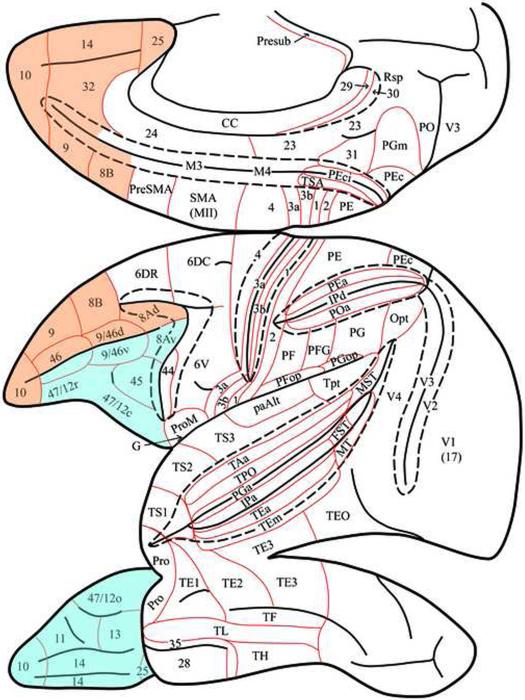
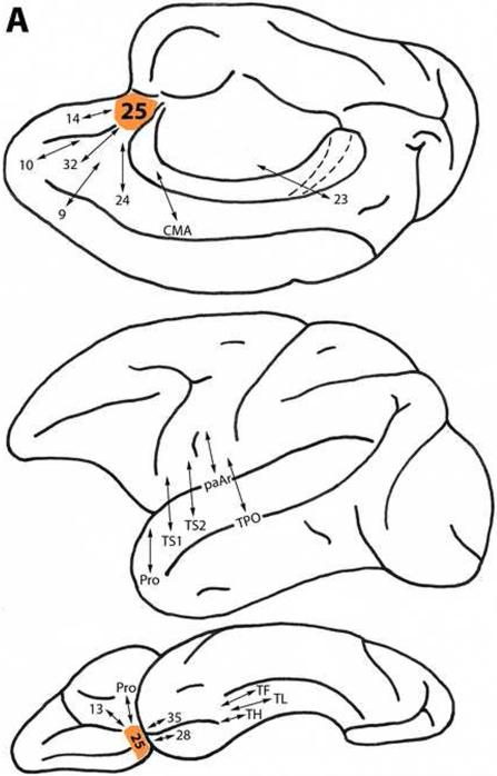
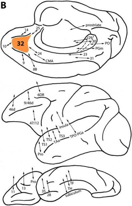
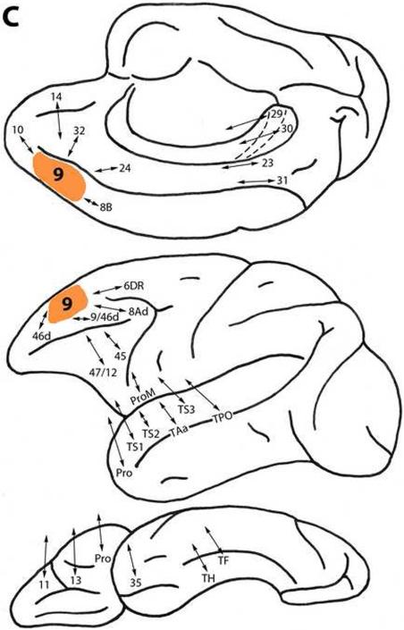
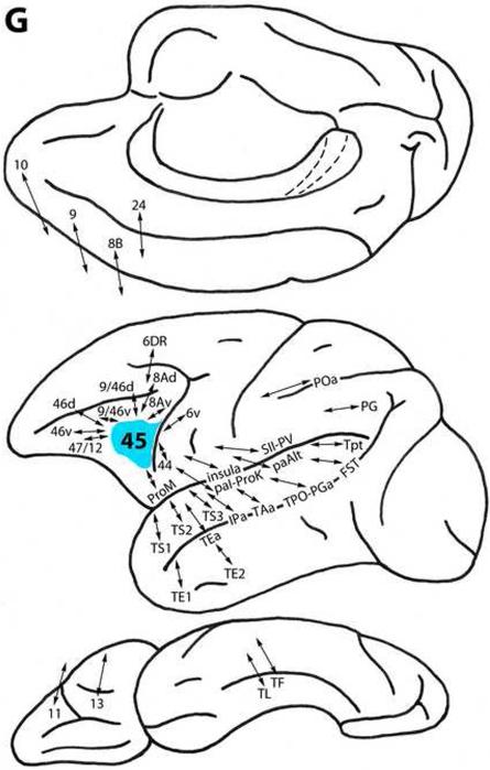
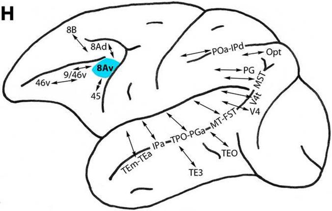
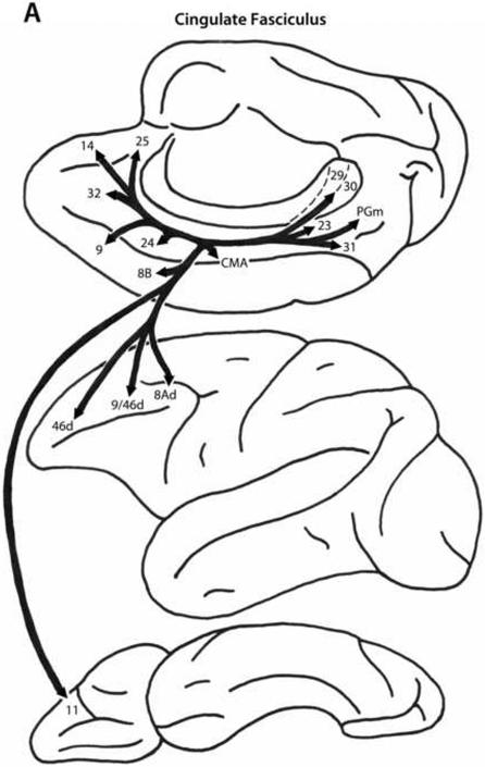
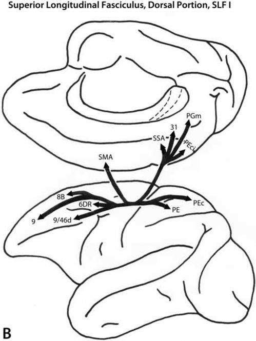
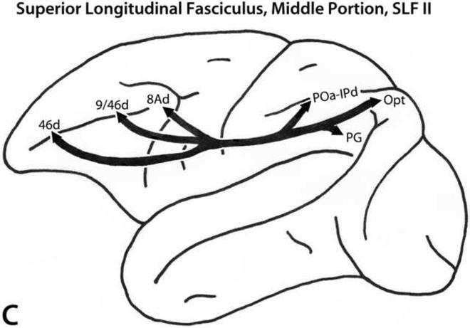
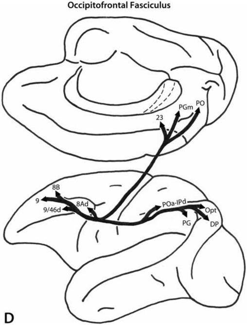
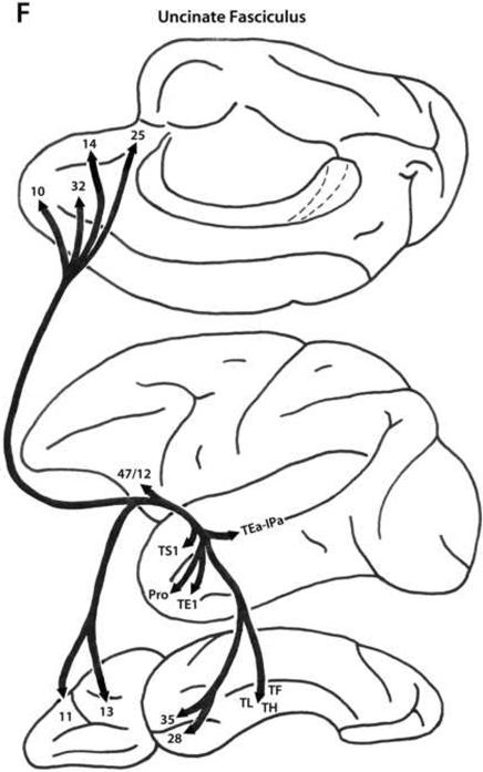
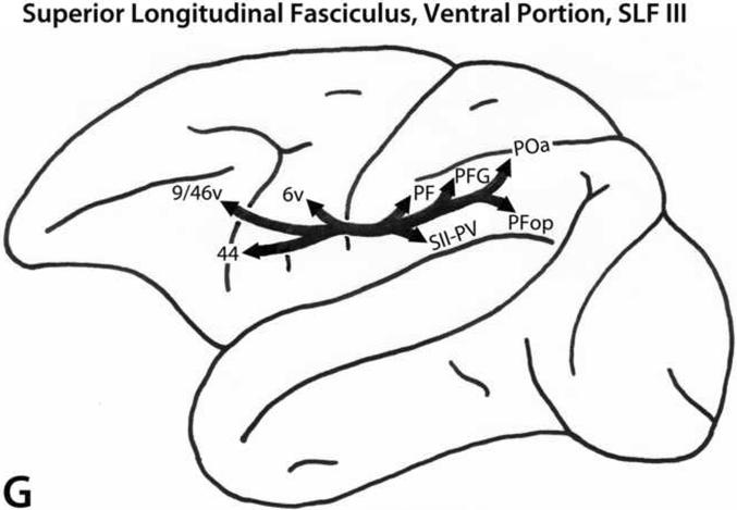
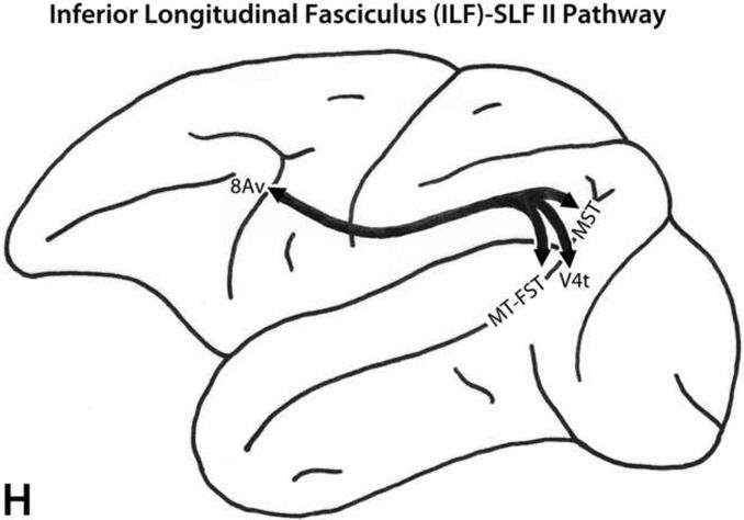
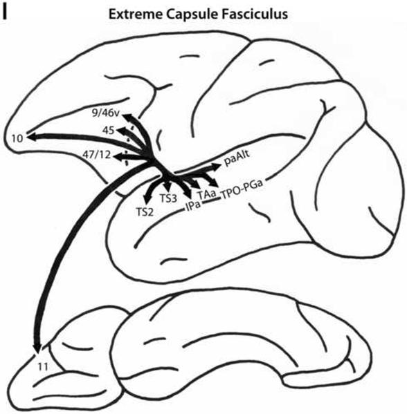
One dimension of understanding the functions of the prefrontal cortex is knowledge of cortical connectivity. We have surveyed three aspects of prefrontal cortical connections: local projections (within the frontal lobe), the termination patterns of long association (post-Rolandic) projections, and the trajectories of major fiber pathways. The local connections appear to be organized in relation to dorsal (hippocampal origin) and ventral (paleocortical origin) architectonic trends. According to the proposal of a dual origin of the cerebral cortex, cortical areas can be traced as originating from archicortex (hippocampus) on the one hand, and paleocortex, on the other hand, in a stepwise manner (e.g., Sanides, 1969; Pandya and Yeterian, 1985). Prefrontal areas within each trend are connected with less architectonically differentiated areas, and also with more differentiated areas. Such organization may allow for the systematic exchange of information within each architectonic trend. The long connections of the prefrontal cortex with post-Rolandic regions seem to be organized preferentially in relation to dorsal and ventral prefrontal architectonic trends. Prefrontal areas are connected with post-Rolandic auditory, visual and somatosensory association areas, and with multimodal and paralimbic regions. This long connectivity likely works in conjunction with local connections to serve prefrontal cortical functions. The afferent and efferent connections of the prefrontal cortex with post-Rolandic regions are conveyed by specific long association pathways. These pathways as well appear to be organized in relation to dorsal and ventral prefrontal architectonic trends. Finally, although prefrontal areas have preferential connections in relation to dual architectonic trends, it is clear that there are interconnections between and among areas in each trend, which may provide a substrate for the overall integrative function of the prefrontal cortex. Prefrontal corticocortical connectivity may help to elucidate both region-specific and integrative perspectives on the functions of the prefrontal cortex.


Figures

Figure 1
Figure 1
Schematic representations of the medial, lateral, and ventral surfaces of the cerebral cortex of the rhesus monkey (Macaca mulatta) according to Petrides and Pandya (1994, 2007). The architectonic areas depicted in these diagrams are based on the findings of several investigators: occipital (Paxinos et al., 2000), parietal (Pandya and Seltzer, 1982), inferotemporal and superior temporal sulcal (Seltzer and Pandya, 1978; Ungerleider and Desimone, 1986), superior temporal gyrus and supratemporal plane (Pandya and Sanides, 1973; Krubitzer and Kaas, 1990); insular, parietotemporal opercular and frontotemporal opercular (Jones and Burton, 1976; Mesulam and Mufson, 1982; Krubitzer and Kaas, 1990; Krubitzer et al., 1995), premotor (Barbas and Pandya, 1987), prefrontal (Petrides and Pandya, 1994), cingulate (Vogt et al., 1987), and parahippocampal (Blatt et al., 2003) regions. Areas of the dorsal prefrontal (archicortical) architectonic trend are shown in orange, and those of the ventral prefrontal (paleocortical) architectonic trend are shown in blue.
Figure 2
Figure 2
A. Diagrammatic representations of the medial, lateral and ventral surfaces of the macaque monkey cerebral hemisphere showing bidirectional local (within the frontal lobe) and long (post-Rolandic) association connections of areas (shown in orange) in the dorsal prefrontal architectonic trend. A: Area 25. B: Area 32. C: Area 9. D: Area 8B. E: Area 10. F: Area 46d. G: Area 9/46d. H: Area 8Ad. Note that in these diagrams and in those in Figure 3, the arrows indicate the connectivity of the different prefrontal areas of interest and not the specific trajectory of those connections.
Figure 2
Figure 2
A. Diagrammatic representations of the medial, lateral and ventral surfaces of the macaque monkey cerebral hemisphere showing bidirectional local (within the frontal lobe) and long (post-Rolandic) association connections of areas (shown in orange) in the dorsal prefrontal architectonic trend. A: Area 25. B: Area 32. C: Area 9. D: Area 8B. E: Area 10. F: Area 46d. G: Area 9/46d. H: Area 8Ad. Note that in these diagrams and in those in Figure 3, the arrows indicate the connectivity of the different prefrontal areas of interest and not the specific trajectory of those connections.
Figure 3
Figure 3
A. Diagrammatic representations of the medial, lateral and ventral surfaces of the macaque monkey cerebral hemisphere showing bidirectional local (within the frontal lobe) and long (Post-Rolandic) association connections of areas (shown in blue) in the ventral prefrontal architectonic trend. A: Area 13. B: Area 14. C: Area 11. D: Area 47/12. E: Area 46v. F: Area 9/46v. G: Area 45. H: Area 8Av.
Figure 3
Figure 3
A. Diagrammatic representations of the medial, lateral and ventral surfaces of the macaque monkey cerebral hemisphere showing bidirectional local (within the frontal lobe) and long (Post-Rolandic) association connections of areas (shown in blue) in the ventral prefrontal architectonic trend. A: Area 13. B: Area 14. C: Area 11. D: Area 47/12. E: Area 46v. F: Area 9/46v. G: Area 45. H: Area 8Av.
Figure 4
Figure 4
A. Schematic diagrams of the medial, lateral and ventral surfaces of the macaque monkey cerebral hemisphere depicting the trajectories of the long prefrontal fiber pathways. A: Cingulate fasciculus (dorsal trend) limbic pathway. B: Superior longitudinal fasciculus, dorsal portion (SLF I, dorsal trend) somatosensory pathway. C: Superior longitudinal fasciculus, middle portion (SLF II, dorsal trend) multimodal pathway. D: Occipitofrontal (dorsal trend) visual pathway. E: Arcuate fasciculus (dorsal trend) auditory pathway. F: Uncinate fasciculus (ventral trend) limbic pathway. G: Superior longitudinal fasciculus, ventral portion (SLF III, ventral trend) somatosensory pathway. H: Inferior longitudinal fasciculus - superior longitudinal fasciculus, middle portion (ILF - SLF II, ventral trend) visual pathway. I: Extreme capsule fasciculus (ventral trend) auditory and multimodal pathway. Note that the arrows and their trajectories indicate connectivity between areas, and do not represent the magnitude of those connectional relationships. For more detailed descriptions of these pathways, see Schmahmann and Pandya (2006).
Figure 4
Figure 4
A. Schematic diagrams of the medial, lateral and ventral surfaces of the macaque monkey cerebral hemisphere depicting the trajectories of the long prefrontal fiber pathways. A: Cingulate fasciculus (dorsal trend) limbic pathway. B: Superior longitudinal fasciculus, dorsal portion (SLF I, dorsal trend) somatosensory pathway. C: Superior longitudinal fasciculus, middle portion (SLF II, dorsal trend) multimodal pathway. D: Occipitofrontal (dorsal trend) visual pathway. E: Arcuate fasciculus (dorsal trend) auditory pathway. F: Uncinate fasciculus (ventral trend) limbic pathway. G: Superior longitudinal fasciculus, ventral portion (SLF III, ventral trend) somatosensory pathway. H: Inferior longitudinal fasciculus - superior longitudinal fasciculus, middle portion (ILF - SLF II, ventral trend) visual pathway. I: Extreme capsule fasciculus (ventral trend) auditory and multimodal pathway. Note that the arrows and their trajectories indicate connectivity between areas, and do not represent the magnitude of those connectional relationships. For more detailed descriptions of these pathways, see Schmahmann and Pandya (2006).

References

    1. Andersen RA, Asanuma C, Cowan WM. Callosal and prefrontal associational projecting cell populations in area 7A of the macaque monkey: A study using retrogradely transported fluorescent dyes. Journal of Comparative Neurology. 1985;232(4):443–455. - PubMed
    1. Andersen RA, Asanuma C, Essick G, Siegel RM. Corticocortical connections of anatomically and physiologically defined subdivisions within the inferior parietal lobule. Journal of Comparative Neurology. 1990;296(1):65–113. - PubMed
    1. Arikuni T, Watanabe K, Kubota K. Connections of area 8 with area 6 in the brain of the macaque monkey. Journal of Comparative Neurology. 1988;277(1):21–40. - PubMed
    1. Arikuni T, Sako H, Murata A. Ipsilateral connections of the anterior cingulate cortex with the frontal and medial temporal cortices in the macaque monkey. Neuroscience Research. 1994;21(1):19–39. - PubMed
    1. Bachevalier J, Meunier M, Lu MX, Ungerleider LG. Thalamic and temporal cortex input to medial prefrontal cortex in rhesus monkeys. Experimental Brain Research. 1997;115(3):430–444. - PubMed

Publication types

MeSH terms

LinkOut - more resources